

Smaller Absolute Quantities but Greater Relative Densities of Microvessels Are Associated with Cerebellar Degeneration in Lurcher Mice
- PMID: 27147979
- PMCID: PMC4835681
- DOI: 10.3389/fnana.2016.00035
Smaller Absolute Quantities but Greater Relative Densities of Microvessels Are Associated with Cerebellar Degeneration in Lurcher Mice
Abstract
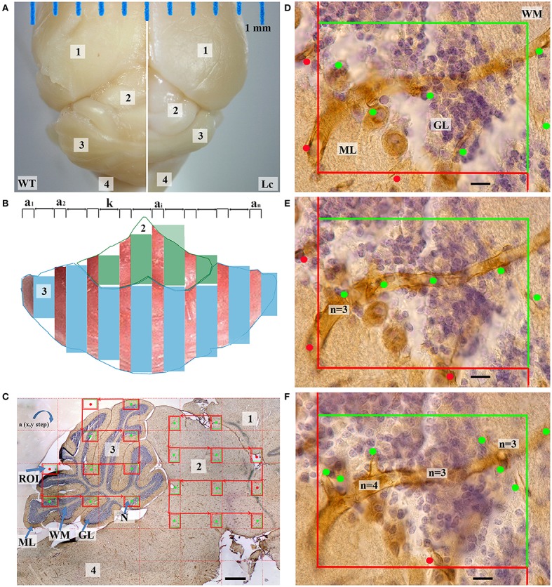
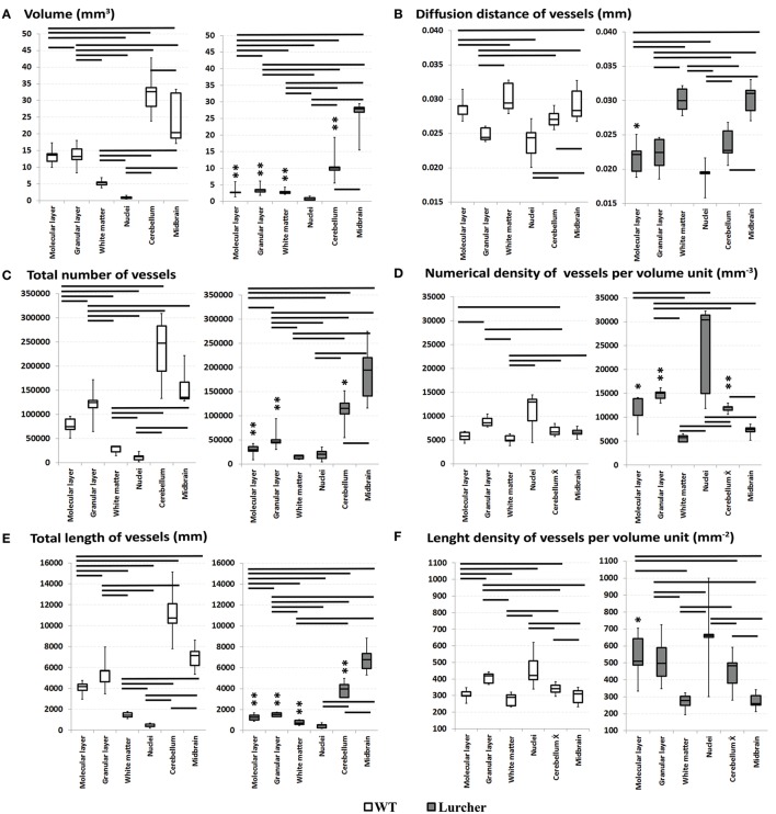
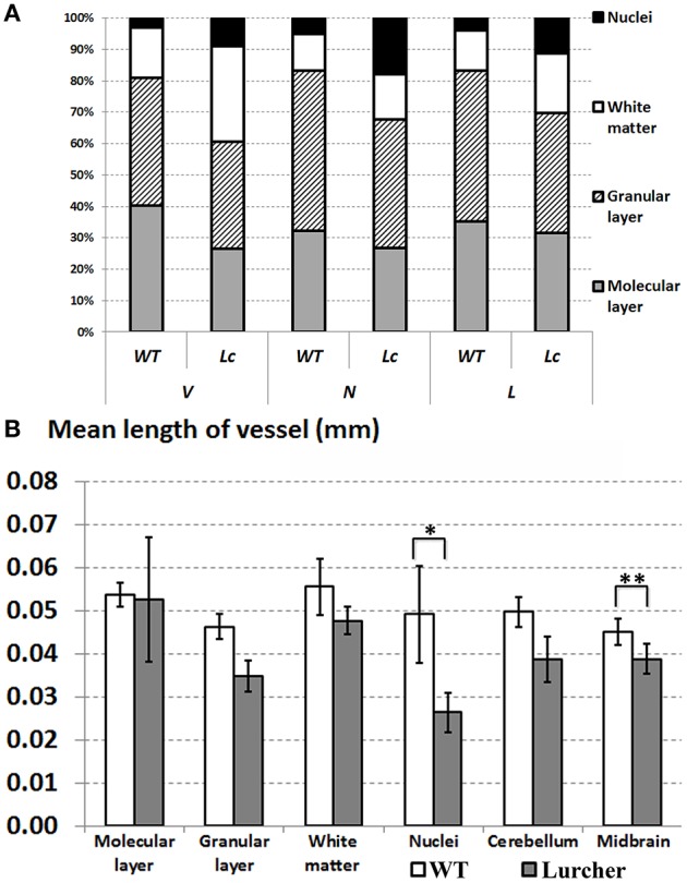
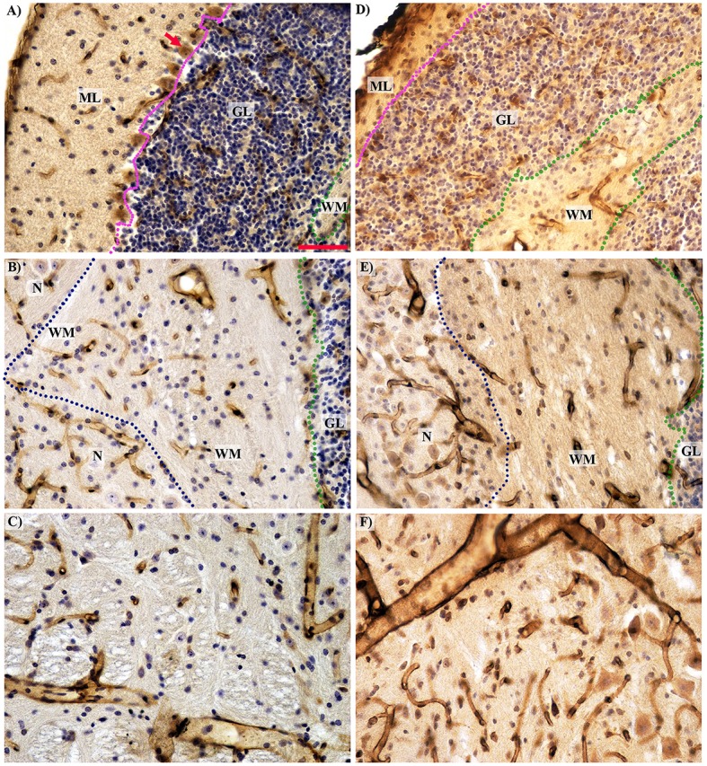
Degenerative affections of nerve tissues are often accompanied by changes of vascularization. In this regard, not much is known about hereditary cerebellar degeneration. In this study, we compared the vascularity of the individual cerebellar components and the mesencephalon of 3-month-old wild type mice (n = 5) and Lurcher mutant mice, which represent a model of hereditary olivocerebellar degeneration (n = 5). Paraformaldehyde-fixed brains were processed into 18-μm thick serial sections with random orientation. Microvessels were visualized using polyclonal rabbit anti-laminin antibodies. Then, the stacks comprised of three 5-μm thick optical sections were recorded using systematic uniform random sampling. Stereological assessment was conducted based on photo-documentation. We found that each of the cerebellar components has its own features of vascularity. The greatest number and length of vessels were found in the granular layer; the number of vessels was lower in the molecular layer, and the lowest number of vessels was observed in the cerebellar nuclei corresponding with their low volume. Nevertheless, the nuclei had the greatest density of blood vessels. The reduction of cerebellum volume in the Lurcher mice was accompanied by a reduction in vascularization in the individual cerebellar components, mainly in the cortex. Moreover, despite the lower density of microvessels in the Lurcher mice compared with the wild type mice, the relative density of microvessels in the cerebellar cortex and nuclei was greater in Lurcher mice. The complete primary morphometric data, in the form of continuous variables, is included as a supplement. Mapping of the cerebellar and midbrain microvessels has explanatory potential for studies using mouse models of neurodegeneration.
Keywords: Lurcher; blood microvessels; cerebellum; cerebral degeneration; laminin; mice; quantitative histology; stereology.
Figures
Similar articles
-
Lurcher Mouse as a Model of Cerebellar Syndromes.Cerebellum. 2025 Feb 28;24(2):54. doi: 10.1007/s12311-025-01810-5. Cerebellum. 2025. PMID: 40016581 Free PMC article. Review.
-
Reduction of Microvessel Number and Length in the Cerebellum of Purkinje Cell Degeneration Mice.Cerebellum. 2024 Apr;23(2):471-478. doi: 10.1007/s12311-023-01556-y. Epub 2023 Apr 18. Cerebellum. 2024. PMID: 37071329
-
Numerical and length densities of microvessels in the human brain: Correlation with preferential orientation of microvessels in the cerebral cortex, subcortical grey matter and white matter, pons and cerebellum.J Chem Neuroanat. 2018 Mar;88:22-32. doi: 10.1016/j.jchemneu.2017.11.005. Epub 2017 Nov 4. J Chem Neuroanat. 2018. PMID: 29113946
-
Motor skills and motor learning in Lurcher mutant mice during aging.Neuroscience. 2001;102(3):615-23. doi: 10.1016/s0306-4522(00)00509-1. Neuroscience. 2001. PMID: 11226698
-
The Lurcher mouse: fresh insights from an old mutant.Brain Res. 2007 Apr 6;1140:4-18. doi: 10.1016/j.brainres.2005.11.086. Epub 2006 Jan 17. Brain Res. 2007. PMID: 16412991 Review.
Cited by
-
Embryonic Cerebellar Graft Morphology Differs in Two Mouse Models of Cerebellar Degeneration.Cerebellum. 2019 Oct;18(5):855-865. doi: 10.1007/s12311-019-01067-9. Cerebellum. 2019. PMID: 31418135
-
Lurcher Mouse as a Model of Cerebellar Syndromes.Cerebellum. 2025 Feb 28;24(2):54. doi: 10.1007/s12311-025-01810-5. Cerebellum. 2025. PMID: 40016581 Free PMC article. Review.
-
Alcohol-Induced Alterations in the Vascular Basement Membrane in the Substantia Nigra of the Adult Human Brain.Biomedicines. 2022 Apr 1;10(4):830. doi: 10.3390/biomedicines10040830. Biomedicines. 2022. PMID: 35453580 Free PMC article.
-
Reduction of Microvessel Number and Length in the Cerebellum of Purkinje Cell Degeneration Mice.Cerebellum. 2024 Apr;23(2):471-478. doi: 10.1007/s12311-023-01556-y. Epub 2023 Apr 18. Cerebellum. 2024. PMID: 37071329
-
Quantification of Solid Embryonic Cerebellar Graft Volume in a Degenerative Ataxia Model.Cerebellum. 2024 Oct;23(5):1811-1823. doi: 10.1007/s12311-024-01676-z. Epub 2024 Mar 2. Cerebellum. 2024. PMID: 38430389
References
LinkOut - more resources
Full Text Sources
Other Literature Sources