. 2016 Apr 12:10:34.
doi: 10.3389/fnsys.2016.00034. eCollection 2016.

Superior Colliculus Responses to Attended, Unattended, and Remembered Saccade Targets during Smooth Pursuit Eye Movements

Affiliations

Superior Colliculus Responses to Attended, Unattended, and Remembered Saccade Targets during Smooth Pursuit Eye Movements

Suryadeep Dash et al. Front Syst Neurosci. .

Abstract

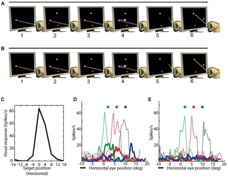
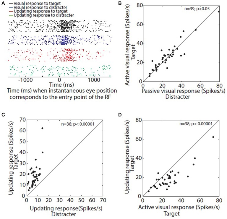
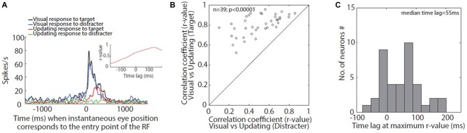
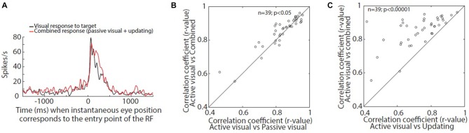
In realistic environments, keeping track of multiple visual targets during eye movements likely involves an interaction between vision, top-down spatial attention, memory, and self-motion information. Recently we found that the superior colliculus (SC) visual memory response is attention-sensitive and continuously updated relative to gaze direction. In that study, animals were trained to remember the location of a saccade target across an intervening smooth pursuit (SP) eye movement (Dash et al., 2015). Here, we modified this paradigm to directly compare the properties of visual and memory updating responses to attended and unattended targets. Our analysis shows that during SP, active SC visual vs. memory updating responses share similar gaze-centered spatio-temporal profiles (suggesting a common mechanism), but updating was weaker by ~25%, delayed by ~55 ms, and far more dependent on attention. Further, during SP the sum of passive visual responses (to distracter stimuli) and memory updating responses (to saccade targets) closely resembled the responses for active attentional tracking of visible saccade targets. These results suggest that SP updating signals provide a damped, delayed estimate of attended location that contributes to the gaze-centered tracking of both remembered and visible saccade targets.

Keywords: attention; saccades; smooth pursuit; spatial updating; superior colliculi.


Figures

Figure 1
Figure 1
Paradigms. (A) Smooth pursuit (SP) visual task: (1) monkey fixated a white dot on the CRT monitor; (2–3) while fixating a peripheral target (white) and distracter (orange) appeared and continued to be visible; (4) the fixation dot started to move with a constant velocity (10°/s). Monkey followed the white dot with SP. The paradigm was configured in such a way that the target or distracter (white or orange dot) corresponded to the visual receptive fields (RF) of the neuron somewhere during the SP; (5) at an unpredictable time during SP the white SP target disappeared; (6) at this point the animal was required to make a saccade towards the visual target (white peripheral dot). (B) SP updating task: (1) monkey fixated a white dot on the CRT monitor; (2) while fixating a peripheral target (white) and distracter (orange) appeared for 200 ms and disappeared; (3) the animal kept the location of target in memory (memory target) and continued looking at fixation dot for another 300–500 ms; (4) the fixation dot started to move with a constant velocity (10°/s). Monkey followed the white dot with SP. The paradigm was configured in such a way that the memory target corresponded to the visual RF of the neuron somewhere during the SP; (5) at an unpredictable time during SP the white dot disappeared; (6) at this point the animal was required to make a saccade towards the memory target. The paradigm required the animals to continuously update its location during the SP. (C) One dimensional visual RF parallel to SP direction: the panel gives the average firing rate for visual stimulation (black) for various targets during memory saccade paradigm. The choice of these targets depended on the configuration of subsequent SP-visual/updating task. The targets corresponds to nine locations spanning from −16 to 16° in horizontal axis with a fixed vertical component of 5°. This visual neuron was most active at 0°/5° (horizontal/vertical), and was also active at 4°/5°. (D) Visual and updating responses to target: shows the neural activity in screen coordinates during SP visual/updating task (neural activity as a function of horizontal eye position). This is an example of updating across horizontal SP (from 10° leftward towards 18° rightwards; black arrow indicates SP direction) followed by a saccade to different visual/memory target (circles; targets were not visible during SP in SP updating task but visible during SP visual task). Colors indicate sets of trials associated with different visual/memory targets. (E) Visual and updating responses to distracter: same as (D) except visual and updating responses were collected when distracter corresponded with the RF of the neuron.
Figure 2
Figure 2
(A) Time aligned raster display of visual and updating response: black and blue raster’s corresponds to visual responses when target or distracter corresponded with entry point of the neuron’s RF, respectively. Red and green raster’s corresponds to updating responses for target and distracter, respectively. (B) Comparison of visual response for target and distracter: average visual response for target is plotted as a function of visual response for distracter. (C) Comparison of updating responses for target and distracter: average updating response for target is plotted as a function of updating response for distracter. All the neurons show a higher updating response for target when compared with that for distracter. (D) Comparison of visual and updating responses for target: average updating response is plotted as a function of average visual response. Almost all the neurons show a higher visual response compared with updating response.
Figure 3
Figure 3
(A) Time aligned spike density function (SDF) of visual and updating responses: black and blue SDF corresponds to visual responses when target or distracter corresponded with entry point of the example neuron’s RF, respectively. Red and green SDF corresponds to updating responses for target and distracter, respectively. The inset shows the correlation between visual and updating response towards the target at different time lags. This neuron shows maximum correlation (r-value) when updating response lags behind the visual response by 137 ms. (B) Correlation between visual and updating responses: correlation between visual and updating response for target is plotted as a function of correlation for the distracter. All the neurons show a higher correlation between visual and updating response for target when compared with that for distracter. (C) Time lag for best correlation: shows the histogram representation of the time lag or lead when visual and updating responses showed maximum correlation across the sample of neurons. The median lag for the population was 55 ms, i.e., updating response lagged behind visual response by 55 ms.
Figure 4
Figure 4
(A) Time aligned SDF of active visual response and combination of passive visual and active updating responses. (B,C) Correlation between active visual response and combined responses (passive visual response and updating response) compared to correlation between active and passive visual response (B) and correlation between active visual response and updating response (C) at zero time lag.
Figure 5
Figure 5
Schematic representation of different sources of visual, memory and updating signals to superior colliculus (SC). Visual signals (red arrows) is mostly contributed by visual cortex and retina but areas lateral intraparietal area (LIP), dorso-lateral prefrontal cortex (DLPFC) and frontal eye fields (FEF) also contributes visual signals to SC. The short term memory information (blue arrows) could reach SC from DLPFC, FEF, LIP or basal ganglia regions or maintained by intrinsic connections within SC. The eye velocity related updating signal (green arrows) could reach SC from various cortical areas (ventral intraparietal (VIP) and medial superior temporal (MST)) and/or cerebellar regions (oculomotor vermis and ventral paraflocculus). The black arrows indicate motor channels from SC to eye muscles through pendunculopontine reticular formation (PPRF) and rostral interstitial nucleus of medial longitudinal fasciculus (riMLF).

Similar articles

Cited by

References

    1. Armstrong K. M., Chang M. H., Moore T. (2009). Selection and maintenance of spatial information by frontal eye field neurons. J. Neurosci. 29, 15621–15629. 10.1523/JNEUROSCI.4465-09.2009 - DOI - PMC - PubMed
    1. Baddeley A. (2003). Working memory: looking back and looking forward. Nat. Rev. Neurosci. 4, 829–839. 10.1038/nrn1201 - DOI - PubMed
    1. Baddeley A. (2012). Working memory: theories, models and controversies. Annu. Rev. Psychol. 63, 1–29. 10.1146/annurev-psych-120710-100422 - DOI - PubMed
    1. Baker J. T., Harper T. M., Snyder L. H. (2003). Spatial memory following shifts of gaze. J. Neurophysiol. 89, 2564–2576. 10.1152/jn.00610.2002 - DOI - PubMed
    1. Blohm G., Missal M., Lefèvre P. (2005). Processing of retinal and extraretinal signals for memory-guided saccades during smooth pursuit. J. Neurophysiol. 93, 1510–1522. 10.1152/jn.00543.2004 - DOI - PubMed

LinkOut - more resources