Novel Radiofrequency Ablation Strategies for Terminating Atrial Fibrillation in the Left Atrium: A Simulation Study
- PMID: 27148061
- PMCID: PMC4828663
- DOI: 10.3389/fphys.2016.00108
Novel Radiofrequency Ablation Strategies for Terminating Atrial Fibrillation in the Left Atrium: A Simulation Study
Abstract
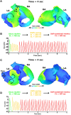
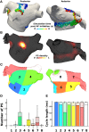
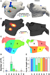
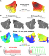
Pulmonary vein isolation (PVI) with radiofrequency ablation (RFA) is the cornerstone of atrial fibrillation (AF) therapy, but few strategies exist for when it fails. To guide RFA, phase singularity (PS) mapping locates reentrant electrical waves (rotors) that perpetuate AF. The goal of this study was to test existing and develop new RFA strategies for terminating rotors identified with PS mapping. It is unsafe to test experimental RFA strategies in patients, so they were evaluated in silico using a bilayer computer model of the human atria with persistent AF (pAF) electrical (ionic) and structural (fibrosis) remodeling. pAF was initiated by rapidly pacing the right (RSPV) and left (LSPV) superior pulmonary veins during sinus rhythm, and rotor dynamics quantified by PS analysis. Three RFA strategies were studied: (i) PVI, roof, and mitral lines; (ii) circles, perforated circles, lines, and crosses 0.5-1.5 cm in diameter/length administered near rotor locations/pathways identified by PS mapping; and (iii) 4-8 lines streamlining the sequence of electrical activation during sinus rhythm. As in pAF patients, 2 ± 1 rotors with cycle length 185 ± 4 ms and short PS duration 452 ± 401 ms perpetuated simulated pAF. Spatially, PS density had weak to moderate positive correlations with fibrosis density (RSPV: r = 0.38, p = 0.35, LSPV: r = 0.77, p = 0.02). RFA PVI, mitral, and roof lines failed to terminate pAF, but RFA perforated circles and lines 1.5 cm in diameter/length terminated meandering rotors from RSPV pacing when placed at locations with high PS density. Similarly, RFA circles, perforated circles, and crosses 1.5 cm in diameter/length terminated stationary rotors from LSPV pacing. The most effective strategy for terminating pAF was to streamline the sequence of activation during sinus rhythm with >4 RFA lines. These results demonstrate that co-localizing 1.5 cm RFA lesions with locations of high PS density is a promising strategy for terminating pAF rotors. For patients immune to PVI, roof, mitral, and PS guided RFA strategies, streamlining patient-specific activation sequences during sinus rhythm is a robust but challenging alternative.
Keywords: ablation; computer modeling; fibrosis; persistent atrial fibrillation; phase singularity mapping.
Figures


Similar articles
-
Focal Impulse and Rotor Modulation for the Treatment of Atrial Fibrillation: Locations and 1 Year Outcomes of Human Rotors Identified Using a 64-Electrode Basket Catheter.J Cardiovasc Electrophysiol. 2017 Apr;28(4):367-374. doi: 10.1111/jce.13157. Epub 2017 Feb 7. J Cardiovasc Electrophysiol. 2017. PMID: 28039924
-
Intermediate term outcome after electrogram guided segmental ostial pulmonary vein isolation using an 8 mm tip catheter for paroxysmal atrial fibrillation.Indian Heart J. 2019 Sep-Oct;71(5):381-386. doi: 10.1016/j.ihj.2019.11.258. Epub 2019 Dec 6. Indian Heart J. 2019. PMID: 32035520 Free PMC article.
-
[Comparison of the effectiveness of pulmonary veins isolation vs linear radiofrequency ablation in paroxysmal atrial fibrillation patients using either mathematical scanning or clinical approach].Kardiologiia. 2012;52(7):50-5. Kardiologiia. 2012. PMID: 22839714 Russian.
-
[Interventional therapy of atrial fibrillation: possibilities and limitations].Dtsch Med Wochenschr. 2010 Mar;135 Suppl 2:S48-54. doi: 10.1055/s-0030-1249209. Epub 2010 Mar 10. Dtsch Med Wochenschr. 2010. PMID: 20221979 Review. German.
-
Catheter Ablation for Long-Standing Persistent Atrial Fibrillation.Methodist Debakey Cardiovasc J. 2015 Apr-Jun;11(2):87-93. doi: 10.14797/mdcj-11-2-87. Methodist Debakey Cardiovasc J. 2015. PMID: 26306125 Free PMC article. Review.
Cited by
-
Variability in pulmonary vein electrophysiology and fibrosis determines arrhythmia susceptibility and dynamics.PLoS Comput Biol. 2018 May 24;14(5):e1006166. doi: 10.1371/journal.pcbi.1006166. eCollection 2018 May. PLoS Comput Biol. 2018. PMID: 29795549 Free PMC article.
-
A Computational Framework to Benchmark Basket Catheter Guided Ablation in Atrial Fibrillation.Front Physiol. 2018 Sep 21;9:1251. doi: 10.3389/fphys.2018.01251. eCollection 2018. Front Physiol. 2018. PMID: 30298012 Free PMC article.
-
Patient-Specific Identification of Atrial Flutter Vulnerability-A Computational Approach to Reveal Latent Reentry Pathways.Front Physiol. 2019 Jan 14;9:1910. doi: 10.3389/fphys.2018.01910. eCollection 2018. Front Physiol. 2019. PMID: 30692934 Free PMC article.
-
Using personalized computer models to custom-tailor ablation procedures for atrial fibrillation patients: are we there yet?Expert Rev Cardiovasc Ther. 2017 May;15(5):339-341. doi: 10.1080/14779072.2017.1317593. Epub 2017 Apr 17. Expert Rev Cardiovasc Ther. 2017. PMID: 28395557 Free PMC article. No abstract available.
-
Commentary: Atrial Fibrillation Dynamics and Ionic Block Effects in Six Heterogeneous Human 3D Virtual Atria with Distinct Repolarization Dynamics.Front Bioeng Biotechnol. 2017 Oct 6;5:59. doi: 10.3389/fbioe.2017.00059. eCollection 2017. Front Bioeng Biotechnol. 2017. PMID: 29057224 Free PMC article. No abstract available.
References
-
- Allessie M. A., Bonke F. I., Schopman F. J. (1977). Circus movement in rabbit atrial muscle as a mechanism of tachycardia. iii. the “leading circle” concept: a new model of circus movement in cardiac tissue without the involvement of an anatomical obstacle. Circ. Res. 41, 9–18. - PubMed
-
- Calkins H., Kuck K. H., Cappato R., Brugada J., Camm A. J., Chen S.-A., et al. . (2012). 2012 HRS/EHRA/ECAS expert consensus statement on catheter and surgical ablation of atrial fibrillation: recommendations for patient selection, procedural techniques, patient management and follow-up, definitions, endpoints, and research trial design: a report of the Heart Rhythm Society (HRS) Task Force on Catheter and Surgical Ablation of Atrial Fibrillation. Developed in partnership with the European Heart Rhythm Association (EHRA), a registered branch of the European Society of Cardiology (ESC) and the European Cardiac Arrhythmia Society (ECAS); and in collaboration with the American College of Cardiology (ACC), American Heart Association (AHA), the Asia Pacific Heart Rhythm Society (APHRS), and the Society of Thoracic Surgeons (STS). Endorsed by the governing bodies of the American College of Cardiology Foundation, the American Heart Association, the European Cardiac Arrhythmia Society, the European Heart Rhythm Association, the Society of Thoracic Surgeons, the Asia Pacific Heart Rhythm Society, and the Heart Rhythm Society. Heart Rhythm 9, 632–696.e21. 10.1016/j.hrthm.2011.12.016 - DOI - PubMed
LinkOut - more resources
Full Text Sources
Other Literature Sources
Research Materials

