Review
. 2016 Apr 12:7:117.
doi: 10.3389/fimmu.2016.00117. eCollection 2016.

The Origin and Activities of IgA1-Containing Immune Complexes in IgA Nephropathy

Affiliations
Review

The Origin and Activities of IgA1-Containing Immune Complexes in IgA Nephropathy

Barbora Knoppova et al. Front Immunol. .

Abstract

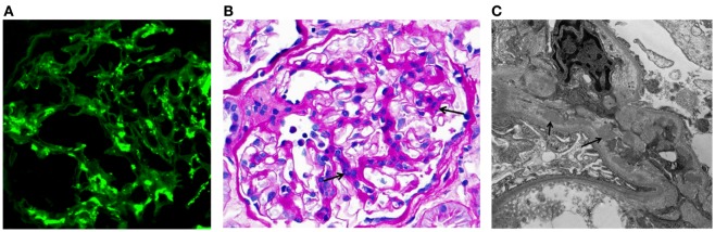
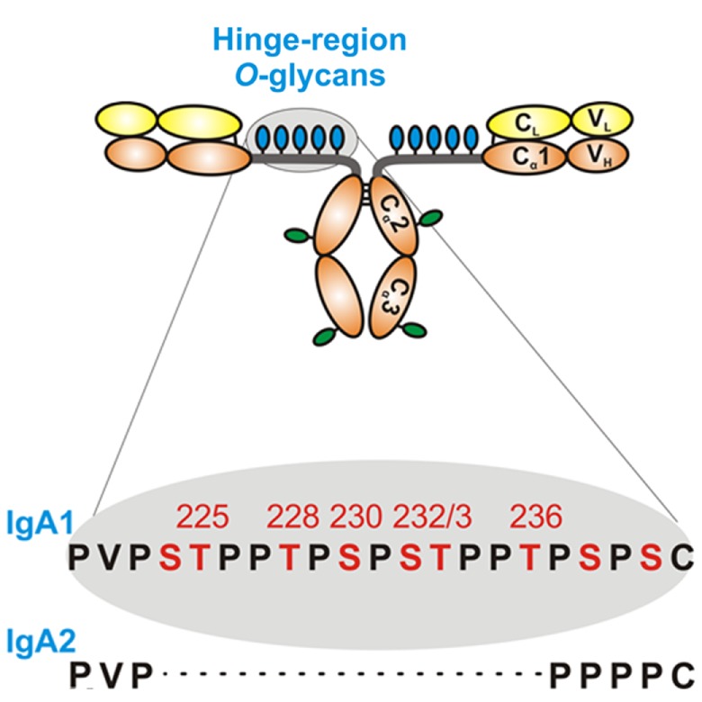
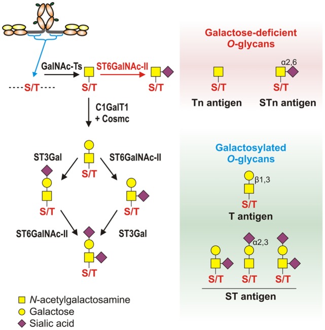
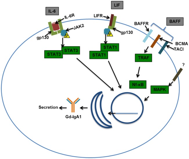
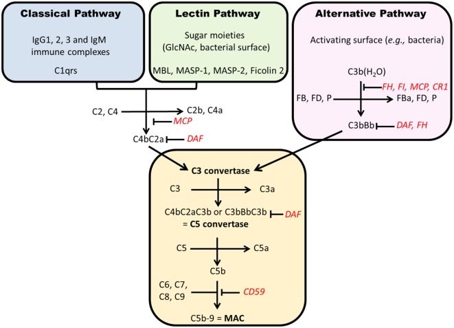
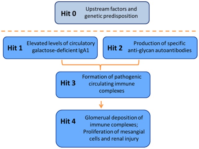
IgA nephropathy (IgAN) is the most common primary glomerulonephritis, frequently leading to end-stage renal disease, as there is no disease-specific therapy. IgAN is diagnosed from pathological assessment of a renal biopsy specimen based on predominant or codominant IgA-containing immunodeposits, usually with complement C3 co-deposits and with variable presence of IgG and/or IgM. The IgA in these renal deposits is galactose-deficient IgA1, with less than a full complement of galactose residues on the O-glycans in the hinge region of the heavy chains. Research from the past decade led to the definition of IgAN as an autoimmune disease with a multi-hit pathogenetic process with contributing genetic and environmental components. In this process, circulating galactose-deficient IgA1 (autoantigen) is bound by antiglycan IgG or IgA (autoantibodies) to form immune complexes. Some of these circulating complexes deposit in glomeruli, and thereby activate mesangial cells and induce renal injury through cellular proliferation and overproduction of extracellular matrix components and cytokines/chemokines. Glycosylation pathways associated with production of the autoantigen and the unique characteristics of the corresponding autoantibodies in patients with IgAN have been uncovered. Complement likely plays a significant role in the formation and the nephritogenic activities of these complexes. Complement activation is mediated through the alternative and lectin pathways and probably occurs systemically on IgA1-containing circulating immune complexes as well as locally in glomeruli. Incidence of IgAN varies greatly by geographical location; the disease is rare in central Africa but accounts for up to 40% of native-kidney biopsies in eastern Asia. Some of this variation may be explained by genetically determined influences on the pathogenesis of the disease. Genome-wide association studies to date have identified several loci associated with IgAN. Some of these loci are associated with the increased prevalence of IgAN, whereas others, such as deletion of complement factor H-related genes 1 and 3, are protective against the disease. Understanding the molecular mechanisms and genetic and biochemical factors involved in formation and activities of pathogenic IgA1-containing immune complexes will enable the development of future disease-specific therapies as well as identification of non-invasive disease-specific biomarkers.

Keywords: IgA; autoantibodies; complement C3; immune complexes; nephropathy.


Figures

Figure 1
Figure 1
Examples of immunofluorescence-, light-, and electron-microscopy features of renal biopsy specimens from patients with IgAN. (A) Immunofluorescence staining for IgA in a kidney biopsy specimen from a patient with IgAN showing mesangial staining. (B) Periodic acid–Schiff staining of a kidney biopsy specimen from a patient with IgAN. Arrows indicate mesangial expansion and hypercellularity. (C) Electron micrograph of kidney biopsy specimen from a patient with IgAN. Arrows point to examples of electron-dense material representative of mesangial and paramesangial immune complex deposits. Images are courtesy of Dr. Huma Fatima (B,C) and Dr. Lea Novak (A), Department of Pathology, UAB.
Figure 2
Figure 2
Hinge-region glycosylation of human IgA1 and comparison of amino-acid sequences of human IgA1 and IgA2. Human IgA1 has nine Ser (S) and Thr (T) amino-acid residues in the hinge-region segment (between constant regions C1 and C2 of the heavy chains). Usually, three to six clustered O-glycans are attached per hinge region. IgA2 hinge region is shorter compared to that of IgA1, does not have Ser and Thr residues and, thus, IgA2 does not have O-glycans. Moreover, each IgA1 heavy chain has two N-glycans, one in the C2 domain and the second in the tailpiece portion of the C3 domain.
Figure 3
Figure 3
Pathways of O-glycosylation of IgA1 hinge region, including galactose-deficient and galactosylated O-glycans. Left panel: O-glycosylation of IgA1 hinge region occurs in the Golgi apparatus and begins with attachment of N-acetylgalactosamine (GalNAc) to Ser or Thr by an enzyme of UDP-GalNAc:polypeptide GalNAc-transferases family (GalNAc-Ts). In patients with IgAN, some terminal GalNAc residues may be prematurely sialylated by GalNAc α2,6-sialyltransferase (ST6GalNAc) (red arrow); this step prevents addition of galactose (the glycan thus remains galactose-deficient). In healthy individuals, GalNAc-α-Ser/Thr residue can be normally modified by addition of galactose, catalyzed by UDP-galactose:GalNAc-α-Ser/Thr β1,3-galactosyltransferase (C1GalT1); stability of C1GalT1 requires molecular chaperone Cosmc. Galβ1,3-GalNAc structures may be further modified by addition of sialic acid to galactose residues through the activity of Galβ1,3-GalNAc α2,3-sialyltransferase (ST3Gal) and/or to GalNAc residues through the activity of ST6GalNAc. Right panel: galactose-deficient O-glycans consist of terminal GalNAc, also known as Tn antigen, or GalNAc with α2,6-linked sialic acid, also known as STn antigen. Galactosylated O-glycans are disaccharides consisting of galactose and GalNAc (Galβ1,3-GalNAcα1-O-Ser/Thr, also known as T antigen) and may be modified by sialic acid (also known as ST antigen). T antigen does not carry sialic acid, but ST antigen has sialic acid attached to galactose and/or GalNAc.
Figure 4
Figure 4
Examples of signaling pathways that may affect production of galactose-deficient IgA1. Interleukin-6 (IL-6) binds to the IL-6 receptor (IL-6R) and through co-receptor gp130 (glycoprotein 130) activates Janus kinase 2 (JAK2), which in turn phosphorylates signal transducer and activator transcription 3 (STAT3), leading to its dimerization and nuclear translocation. Leukemia inhibitory factor (LIF) binds gp130 and LIF receptor (LIFR) that also activates JAK2, leading to STAT1/3 activation and nuclear translocation. B-cell-activating factor (BAFF) can bind multiple receptors, BAFF receptor (BR), B-cell maturation antigen (BCMA), and transmembrane activator and calcium-modulating/cyclophilin ligand protein (TACI). Activation of TNF-receptor-associated factor (TRAF) by TACI, BAFFR, and BCMA leads to NF-kB activation and nuclear translocation. Mitogen-activated protein kinase (MAPK) activation has been shown to affect O-glycosylation and thus was included in this scheme. Nuclear translocation of the STATs and NF-kB may be important in driving production of Gd-IgA1 in IgA1-producing cells and/or maintaining viability/proliferation of these cells in patients with IgAN.
Figure 5
Figure 5
Complement activation pathways. Each pathway results in formation of a C3 convertase that, after addition of C3b, becomes a C5 convertase. The generation of C5b starts the formation of membrane attack complex (C5b–9). Regulatory factors are in red. CR1, complement receptor 1; FD, factor D; MAC, membrane attack complex; MCP, membrane cofactor protein; P, properdin; DAF, decay accelerating factor; MBL, mannan-binding lectin; MASP, MBL-associated serine proteases.
Figure 6
Figure 6
C3 proteolytic cascade. The hydrolysis of C3 leads to release of activation products C3a – an anaphylatoxin – and C3b. C3b binds activating surfaces, such as a bacterial cell wall, triggering the alternative pathway cascade. This activation is controlled by regulator molecules, such as FI, FH, and complement receptor 1 (CR1), that degrade C3b into products that cannot contribute to the formation of the C5 convertase (iC3b, C3c, C3dg, and C3d). Detection of these inactive breakdown products is considered evidence of activation of C3. The numbers, in kilodaltons, represent the molecular masses of the corresponding polypeptides. MCP, membrane cofactor protein.
Figure 7
Figure 7
Multi-hit hypothesis for pathogenesis of IgAN. Several processes are involved in development of IgAN. Circulatory Gd-IgA1 (Hit 1) is recognized by Gd-IgA1-specific autoantibodies (Hit 2) that leads to formation of pathogenic Gd-IgA1-containing circulating immune complexes (Hit 3). Some of these complexes reach the renal glomeruli to bind to mesangial cells and activate them, thereby inducing renal injury (Hit 4).

References

    1. Berger J, Hinglais N. [Intercapillary deposits of IgA-IgG]. J Urol Nephrol (Paris) (1968) 74:694–5. - PubMed
    1. Jennette JC. The immunohistology of IgA nephropathy. Am J Kidney Dis (1988) 12:348–52.10.1016/S0272-6386(88)80022-2 - DOI - PubMed
    1. Cattran DC, Coppo R, Cook HT, Feehally J, Roberts IS, Troyanov S, et al. The Oxford classification of IgA nephropathy: rationale, clinicopathological correlations, and classification. Kidney Int (2009) 76:534–45.10.1038/ki.2009.243 - DOI - PubMed
    1. Roberts IS, Cook HT, Troyanov S, Alpers CE, Amore A, Barratt J, et al. The Oxford classification of IgA nephropathy: pathology definitions, correlations, and reproducibility. Kidney Int (2009) 76:546–56.10.1038/ki.2009.168 - DOI - PubMed
    1. Berthoux FC, Mohey H, Afiani A. Natural history of primary IgA nephropathy. Semin Nephrol (2008) 28:4–9.10.1016/j.semnephrol.2007.10.001 - DOI - PubMed