Review
. 2016 Apr 12:4:24.
doi: 10.3389/fcell.2016.00024. eCollection 2016.

The Exocyst Complex in Health and Disease

Affiliations
Review

The Exocyst Complex in Health and Disease

Magdalena Martin-Urdiroz et al. Front Cell Dev Biol. .

Abstract

Exocytosis involves the fusion of intracellular secretory vesicles with the plasma membrane, thereby delivering integral membrane proteins to the cell surface and releasing material into the extracellular space. Importantly, exocytosis also provides a source of lipid moieties for membrane extension. The tethering of the secretory vesicle before docking and fusion with the plasma membrane is mediated by the exocyst complex, an evolutionary conserved octameric complex of proteins. Recent findings indicate that the exocyst complex also takes part in other intra-cellular processes besides secretion. These various functions seem to converge toward defining a direction of membrane growth in a range of systems from fungi to plants and from neurons to cilia. In this review we summarize the current knowledge of exocyst function in cell polarity, signaling and cell-cell communication and discuss implications for plant and animal health and disease.

Keywords: exocyst complex; fungi; mammals; pathogens; plants.


Figures
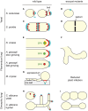
Figure 1
Figure 1
The exocyst complex in fungi. (A,C,E,G,H) Overall localization of the exocyst complex (red and orange) in wild-type cells of representative fungi. For simplicity, S. pombe cells (C,D) are represented as both elongating and dividing. In all fungi, the exocyst localizes at sites of polarized secretion and polarized growth (cyan), the bud tip in budding cells (A,H), cell tip area in tip-growing fungi (C,E) or septal area in dividing cells (A,C). In some cases individual sub-units or sub-complexes are present at different sub-cellular locations, which is only illustrated here for N. crassa (E, red and orange). The exocyst localizes as a ring at the base of the M. oryzae appressorium during rice leaf infection (G). SPK, Spitzenkörper (green). (B,D,F,I) Typical cellular phenotype of exocyst mutants. In the absence of a functional exocyst, most fungal species have a morphological or loss of polarity phenotype, failing to grow a bud (B), growing wider (D), branching out (F), or failing to branch (I). Note that in the case of the hyphal form of C. albicans, three different phenotypes were described. We arbitrarily present here a hypo-branching and globular tip phenotype reported for a sec3 defective mutant. Some fungi also have a cytokinetic defect (D,I). See text for details and references.
Figure 2
Figure 2
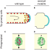
The plant exocyst supports tip-growth and cell division. (A) Tip-growing cells in plants (pollen tubes and root hairs) incorporate membrane and other cargoes delivered via vesicles to the apical growth zone of the PM (cyan). Rapid, rotational streaming of cytoplasm (black arrow) occurs behind the apex and is driven by myosin and actin cables. In pollen tubes SEC6, SEC8, and Exo70 label endomembrane compartments within the apex (orange). In both pollen tubes and root hairs exocyst components decorate the PM in a punctate pattern (red). (B) Loss of exocyst causes shortening and broadening of tip-growing cells. This is potentially consistent with a hypothetical widening of the growth zone (cyan) but could also be caused by alterations in cell wall properties that could result in turgor-driven expansion. (C) Plant cells produce new cell plates (gray) through the action of a cytoskeletal array known as the phragmoplast (microtubule organization within the phragmoplast is depicted in green). The phragmoplast expands from the former site of the spindle and advances toward the outer cell walls where the cell plate will eventually fuse. Exocyst components are enriched at the developing plate during two distinct plate growth phases (red) (Fendrych et al., ; Zhang et al., ; Rybak et al., 2014). (D) Exocyst mutants show a variety of cell plate phenotypes including reduction in the rate of cell plate expansion, increase in the probability of cell plate collapse, and alteration of the content of developing cell walls. See text for details and other references.
Figure 3
Figure 3
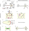
Some of the multiple roles of the animal exocyst complex. (A,C,E,F) Overall localization of the exocyst in physiological conditions (red). For simplicity, the differential localization of individual sub-units is not represented here. The exocyst is mostly present at zones of bulk (leading edge, E), or “finger-like” (neurites, A; cilia, C; filopodia, E) membrane extension. (A) In neurons, some subunits accumulate at dentritic branching points, but not at growing tips of dendrites, whereas others localize to the tip of axon filopodia. (C,E) The exocyst localizes at sites of cell-cell or cell-matrix junctions (tight junctions, TJ, black; cell-cell junctions, CCJ, gray; focal adhesions, FA, green). It was also reported to be associated with trafficking vesicles carrying cell surface receptors (orange), with the migrating basal body (BB), with phagosomes or phagosome-fusing endosomes (purple) and with extra-cellular exosome-like vesicles (ELV, blue). (F) The exocyst can organize as a ring. (B,D,G) Cellular phenotypes associates with a loss of exocyst function. Protrusions necessary for cell function or migration are not formed, cell polarity and tissue integrity are compromised, phagosome acidification is altered which constitutes a window of opportunity for pathogenic bacteria, cells fail to complete cytokinesis, and undergo extra mitoses (nucleus, N). (D) Some toxins secreted by bacteria impair the delivery of the exocyst at cell junctions. See text for details and references.

Similar articles

Cited by

References

    1. Abenza J. F., Couturier E., Dodgson J., Dickmann J., Chessel A., Dumais J., et al. . (2015). Wall mechanics and exocytosis define the shape of growth domains in fission yeast. Nat. Commun. 6, 8400. 10.1038/ncomms9400 - DOI - PMC - PubMed
    1. Ady J. W., Desir S., Thayanithy V., Vogel R. I., Moreira A. L., Downey R. J., et al. . (2014). Intercellular communication in malignant pleural mesothelioma: properties of tunneling nanotubes. Front. Physiol. 5:400. 10.3389/fphys.2014.00400 - DOI - PMC - PubMed
    1. Albeg A., Smith C. J., Chatzigeorgiou M., Feitelson D. G., Hall D. H., Schafer W. R., et al. . (2011). C. elegans multi-dendritic sensory neurons: morphology and function. Mol. Cell. Neurosci. 46, 308–317. 10.1016/j.mcn.2010.10.001 - DOI - PMC - PubMed
    1. Andersen N. J., Yeaman C. (2010). Sec3-containing exocyst complex is required for desmosome assembly in mammalian epithelial cells. Mol. Biol. Cell 21, 152–164. 10.1091/mbc.E09-06-0459 - DOI - PMC - PubMed
    1. Andrews N. W., Almeida P. E., Corrotte M. (2014). Damage control: cellular mechanisms of plasma membrane repair. Trends Cell Biol. 24, 734–742. 10.1016/j.tcb.2014.07.008 - DOI - PMC - PubMed