. 2016 May 5;11(5):e0154805.
doi: 10.1371/journal.pone.0154805. eCollection 2016.

Carboxyl-Terminal SSLKG Motif of the Human Cystinosin-LKG Plays an Important Role in Plasma Membrane Sorting

Affiliations

Carboxyl-Terminal SSLKG Motif of the Human Cystinosin-LKG Plays an Important Role in Plasma Membrane Sorting

Francesco Bellomo et al. PLoS One. .

Abstract

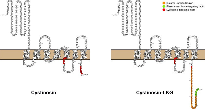
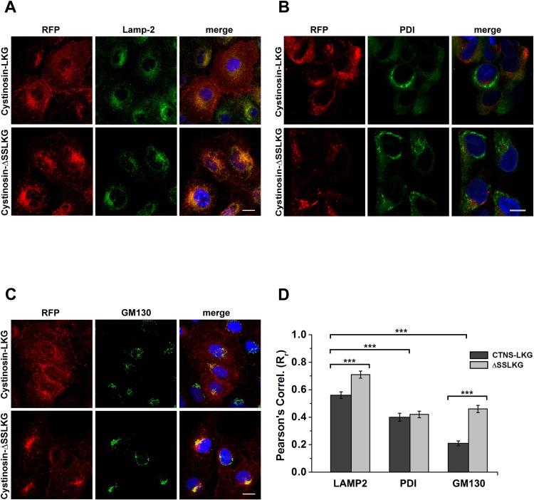
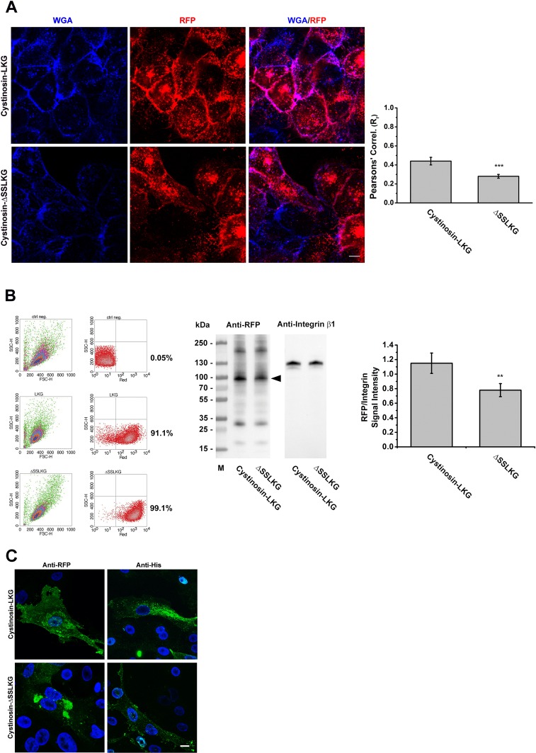
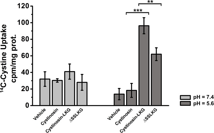
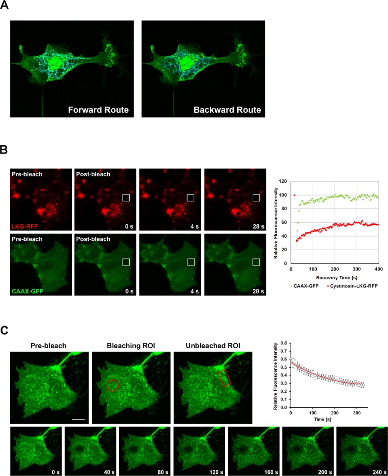
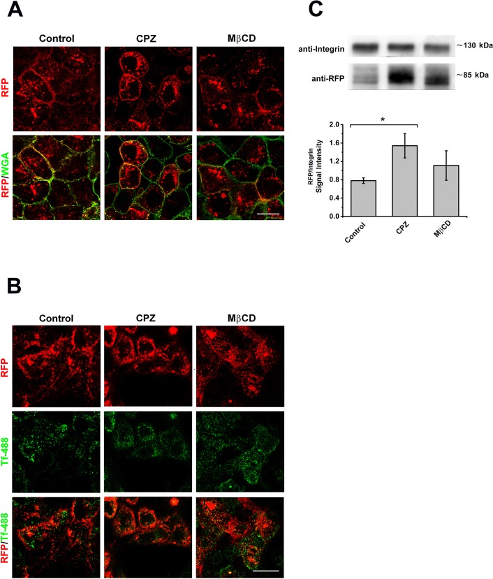
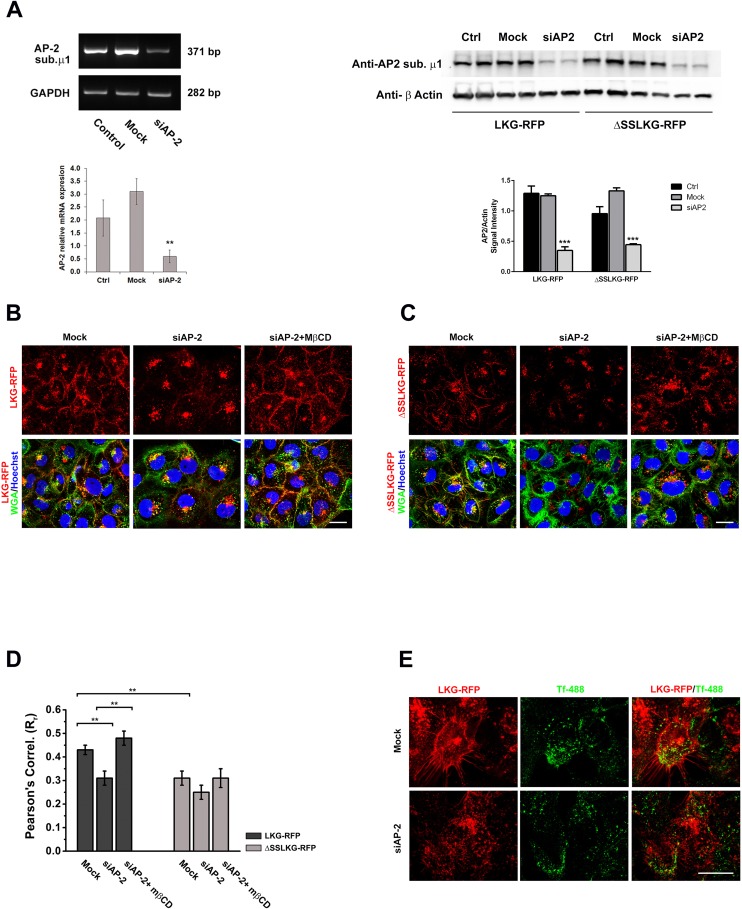
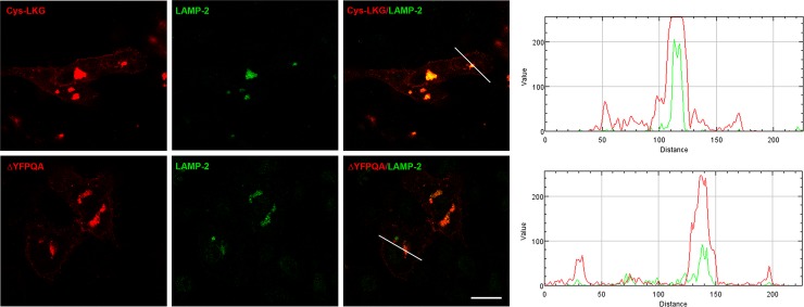
Cystinosin mediates an ATP-dependent cystine efflux from lysosomes and causes, if mutated, nephropathic cystinosis, a rare inherited lysosomal storage disease. Alternative splicing of the last exon of the cystinosin sequence produces the cystinosin-LKG isoform that is characterized by a different C-terminal region causing changes in the subcellular distribution of the protein. We have constructed RFP-tagged proteins and demonstrated by site-directed mutagenesis that the carboxyl-terminal SSLKG sequence of cystinosin-LKG is an important sorting motif that is required for efficient targeting the protein to the plasma membrane, where it can mediate H+ coupled cystine transport. Deletion of the SSLKG sequence reduced cystinosin-LKG expression in the plasma membrane and cystine transport by approximately 30%, and induced significant accumulation of the protein in the Golgi apparatus and in lysosomes. Cystinosin-LKG, unlike the canonical isoform, also moves to the lysosomes by the indirect pathway, after endocytic retrieval from the plasma membrane, mainly by a clathrin-mediated endocytosis. Nevertheless, silencing of AP-2 triggers the clathrin-independent endocytosis, showing the complex adaptability of cystinosin-LKG trafficking.


Conflict of interest statement

Competing Interests: The authors have declared that no competing interests exist.

Figures

Fig 1
Fig 1. Scheme of the cystinosin isoforms structure.
Cystinosin (367 aa) on the left and the cystinosin-LKG isoform (400 aa) on the right, are the main known isoforms to date, for which has been described the transport of cystine. The open-source tool for visualization of proteoforms, PROTTER [32], displayed the hypothetical structure of the two isoforms. Red regions are two targeting motifs for the protein sorting to lysosomes: GYDQL located at the C-terminal end, and YFPQA located in the putative fifth inter-transmembrane loop. Cystinosin-LKG differs from the canonical cystinosin in the C-terminal region (orange) while the proposed motif critical for the protein sorting to the plasma membrane (SSLKG) is highlighted in green.
Fig 2
Fig 2. Subcellular distribution of the cystinosin-LKG and ΔSSLKG mutant.
HK-2 cells were stably transfected with RFP-tagged cystinosin-LKG or with its mutated form, deleted in C-terminal tail for the last five amino acids (ΔSSLKG). Cells were immunolabeled with LAMP-2 for lysosomes (A), PDI for endoplasmic reticulum (B), GM130 for Golgi (C). Scale bar = 10 μm. Analysis of ROIs (Regions of Interest) shows a Pearson’s Correlation (Rr) for RFP with LAMP-2 greater than with other organelle markers (p < 0.0005). In particular, the mutant ΔSSLKG shows an Rr for LAMP-2 significantly increased (p < 0.0005) compared to the wild type cystinosin-LKG. The presence of cystinosin-LKG and ΔSSLKG mutant on ER is low, moreover the analysis shows very low expression of cystinosin-LKG in the Golgi apparatus, whereas high Rr for GM130 in ΔSSLKG mutant suggests that it is accumulated significantly (p < 0.00001) on the Golgi apparatus (D). Means ± SEM of three experiments are shown.
Fig 3
Fig 3. Differential expression of the cystinosin-LKG and ΔSSLKG mutant on the plasma membrane.
HK-2 cells stably transfected with RFP-tagged cystinosin-LKG or ΔSSLKG were stained with WGA Blue to mark the plasma membrane. Colocalization analysis shows that deletion of -SSLKG motif reduces about 35% (p < 0.001) expression of the carrier on the plasma membrane (A). Scale bar = 10 μm, means ± SEM of four experiments are shown. Stably transfected cells with cystinosin-LKG or ΔSSLKG mutant were analyzed by cytofluorimetry to define the transfection efficiency, and a cell surface protein biotinylation for SDS-PAGE analysis was performed. Quantitative analysis by evaluation of RFP signal normalized with Integrin β1 shows about 30% reduction (p < 0.01) in ΔSSLKG mutant (B). Means ± SEM of three experiments are shown. HK-2 cells transiently transfected with RFP- or V5His-tagged cystinosin-LKG and ΔSSLKG were immunolabeled using anti-RFP/ Alexa Fluor® 488 goat anti-rabbit IgG antibody or anti-His Tag/Alexa Fluor® 488 goat anti-mouse IgG antibody, respectively (C). Scale bar = 10 μm.
Fig 4
Fig 4. 14C-cystine uptake from extracellular milieu in HK-2 transfected with different cystinosin isoforms.
14C-cystine uptake assay performed in HK-2 cells transfected with vehicle, cystinosin, cystinosin-LKG, or ΔSSLKG mutant, shows low uptake of radiolabelled L-[14C]-cystine across plasma membrane in absence of a proton gradient (light grey columns). In the presence of a proton gradient (dark grey columns) HK-2 cells overexpressing cystinosin-LKG show a significant 14C-cystine uptake (p < 0.0005) compared to cells transfected with cystinosin; while HK-2 cells overexpressing ΔSSLKG mutant show lower 14C-cystine uptake (p < 0.004) compared to cells transfected with cystinosin-LKG. Means ± SEM of three experiments are shown.
Fig 5
Fig 5. Tracking, FRAP and FLIP analysis in HK-2 transfected with cystinosin-LKG.
HK-2 cells were transiently transfected with EGFP tagged cystinosin-LKG. Tracking of fluorescent vesicles with an estimated diameter > 1 micron, showed the centrifugal (left) and centripetal (right) movements (A). Dynamics of cystinosin-LKG on the plasma membrane was investigated by FRAP (Fluorescence Recovery After Photobleaching). HK-2 cells were transiently transfected with RFP tagged cystinosin-LKG and GFP-CAAX domain as unrelated control protein. Pre-bleach, bleach and post-bleach images are shown of a representative FRAP, and the recovery over time of the bleached region of interest (ROI) is graphically represented. Analysis of these data suggests that about 36% of cystinosin-LKG on the plasma membrane is represented by a mobile fraction (the percentage of maximally recovered fluorescence) with a mean half-time of recovery measuring thalf = 92.2 ± 12.6 s, the time in which 50% of the fluorescence in the bleach spot was recovered (B). To verify if cystinosin-LKG moves from the cytoplasm or lateral boundary, a bleach region of interest (oval) was placed in the cytoplasmic region near plasma membrane and a decay of signal (Fluorescence Loss In Photobleaching–FLIP) was observed only on specific portions of the plasma membrane (rectangle) (C), other regions are not influenced by bleaching. Scale bar = 10 μm, Means ± SEM of four experiments are shown.
Fig 6
Fig 6. Effect of endocytosis inhibitors on endosomal sorting of cystinosin-LKG.
HK-2 cells stably transfected with RFP-tagged cystinosin-LKG, after 48h serum starvation, were treated 30’ with 5 μg/ml chlorpromazine (CPZ) a clathrin-dependent endocytosis inhibitor or with 30 μg/ml methyl-β-cyclodextrin (MβCD) which affects clathrin-independent pathway. Qualitative analysis shows the presence of RFP-tagged cystinosin-LKG on the plasma membrane stained with WGA green, but the RFP signal accumulated more in CPZ treated cells (A). Scale bar = 20 μm. In the same experimental conditions, the uptake of Alexa Fluor® 488 transferrin (488-Tf) was assayed in order to confirm the inhibition of clathrin-dependent endocytosis (B). Scale bar = 20 μm. Quantitative analysis, achieved by protein surface biotinylation and SDS-PAGE, shows that CPZ treatment induces a significant increase of cystinosin-LKG presence on the plasma membrane (C). Means ± SEM of three experiments are shown.
Fig 7
Fig 7. Effect of silencing of AP-2 mu chain on endosomal sorting of cystinosin-LKG and ΔSSLKG mutant.
siRNA to human AP-2 mu chain and a scrambled control were transfected into HK-2 cells overexpressing RFP tagged cystinosin-LKG and ΔSSLKG mutant. The analysis of the AP-2 expression by PCR and western blotting show an efficient silencing with a significant transcript reduction (p < 0.005) (A). After 72h silencing of AP-2 mu chain, expression of cystinosin-LKG on the plasma membrane is significantly reduced. Inhibition of the clathrin-independent pathway by treatment with 30 μg/ml methyl-β-cyclodextrin (MβCD) for 30’ combined to AP-2 silencing, permanently prevents endocytic sorting of cystinosin-LKG from the plasma membrane, triggering the accumulation of the protein on the plasma membrane (B). Scale bar = 20 μm. As previously showed, ΔSSLKG mutant is less expressed on the plasma membrane, and this condition is highlighted by AP-2 silencing. After AP-2 silencing, in fact, ΔSSLKG is very low on plasma membrane and the inhibition of the clathrin-independent pathway with MβCD does not affect significantly the distribution (C). Scale bar = 20 μm. Colocalization analysis between RFP and WGA green (plasma membrane) signals shows the Pearson’s Correlation significantly reduced in ΔSSLKG cells (p < 0.001) and in cystinosin-LKG with AP-2 silencing (p < 0.001). In the latter, after MβCD treatment, the Pearson’s Correlation increases about 24% (p < 0.001), while no significant effects is observed in ΔSSLKG mutant (D). Analysis of transferrin uptake indicates that cystinosin-LKG is expressed on the plasma membrane only in cells where AP-2 was not silenced (transferrin internalized) instead silencing of AP-2 associated to the accumulation of transferrin on the plasma membrane, affects negatively the presence of cystinosin-LKG on the plasma membrane (E). Scale bar = 20 μm. Means ± SEM of four (A, B, C, D) or three (E) experiments are shown.
Fig 8
Fig 8. Colocalization studies of cystinosin-LKG carrying the deletion of lysosomal sorting signal YFPQA.
In HK-2 cells, transiently transfected with the pCTNS-LKG-RFP construct carrying the deletion of the (ΔFPQA), the deleted cystinosin-LKG-RFP could be seen on the plasma membrane as well as in lysosomes. The intensity profile, obtained with RGB Profiler, an ImageJ plugin, showed the RFP signal in lysosomes of ΔFPQA reduced compared to the wild type cystinosin-LKG. Scale bar = 20 μm.

Similar articles

Cited by

References

    1. Nakamura M, Yasuda D, Hirota N, Yamamoto T, Yamaguchi S, Shimizu T, et al. Amino acid residues of G-protein-coupled receptors critical for endoplasmic reticulum export and trafficking. Methods Enzymol. 2013;521: 203–216. 10.1016/B978-0-12-391862-8.00011-9 - DOI - PubMed
    1. Seatter MJ, Drummond R, Kanke T, Macfarlane SR, Hollenberg MD, Plevin R. The role of the C-terminal tail in protease-activated receptor-2-mediated Ca2+ signalling, proline-rich tyrosine kinase-2 activation, and mitogen-activated protein kinase activity. Cell Signal. 2004;16: 21–29. - PubMed
    1. Chung JJ, Shikano S, Hanyu Y, Li M. Functional diversity of protein C-termini: more than zipcoding? Trends Cell Biol. 2002;12: 146–150. - PubMed
    1. Town M, Jean G, Cherqui S, Attard M, Forestier L, Whitmore SA, et al. A novel gene encoding an integral membrane protein is mutated in nephropathic cystinosis. Nat Genet. 1998;18: 319–324. - PubMed
    1. Kalatzis V, Cherqui S, Antignac C, Gasnier B. Cystinosin, the protein defective in cystinosis, is a H(+)-driven lysosomal cystine transporter. EMBO J. 2001;20: 5940–5949. - PMC - PubMed

Publication types

MeSH terms

Substances