Hypoxia Inducible Factor 1 Alpha Is Expressed in Germ Cells throughout the Murine Life Cycle
- PMID: 27148974
- PMCID: PMC4858237
- DOI: 10.1371/journal.pone.0154309
Hypoxia Inducible Factor 1 Alpha Is Expressed in Germ Cells throughout the Murine Life Cycle
Abstract
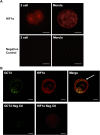
Pluripotent stem cells of the early embryo, and germ line cells, are essential to ensure uncompromised development to adulthood as well as species propagation, respectively. Recently, the transcription factor hypoxia inducible factor 1 alpha (Hif1α) has been shown to have important roles in embryonic stem cells; in particular, regulation of conversion to glycolytic metabolism and, as we have shown, maintenance of functional levels of telomerase. In the present study, we sought to assess whether Hif1α was also expressed in the primitive cells of the murine embryo. We observed expression of Hif1α in pre-implantation embryos, specifically the 2-cell stage, morula, and blastocyst. Robust Hif1α expression was also observed in male and female primordial germ cells. We subsequently assessed whether Hif1α was expressed in adult male and female germ cells. In the testis, Hif1α was robustly expressed in spermatogonial cells, in both juvenile (6-week old) and adult (3-month old) males. In the ovaries, Hif1α was expressed in mature oocytes from adult females, as assessed both in situ and in individual oocytes flushed from super-ovulated females. Analysis of Hif1α transcript levels indicates a mechanism of regulation during early development that involves stockpiling of Hif1α protein in mature oocytes, presumably to provide protection from hypoxic stress until the gene is re-activated at the blastocyst stage. Together, these observations show that Hif1α is expressed throughout the life-cycle, including both the male and female germ line, and point to an important role for Hif1α in early progenitor cells.
Conflict of interest statement
Figures


Similar articles
-
RNAi screen for telomerase reverse transcriptase transcriptional regulators identifies HIF1alpha as critical for telomerase function in murine embryonic stem cells.Proc Natl Acad Sci U S A. 2010 Aug 3;107(31):13842-7. doi: 10.1073/pnas.0913834107. Epub 2010 Jul 19. Proc Natl Acad Sci U S A. 2010. PMID: 20643931 Free PMC article.
-
Murine polo like kinase 1 gene is expressed in meiotic testicular germ cells and oocytes.Mol Reprod Dev. 1995 Aug;41(4):407-15. doi: 10.1002/mrd.1080410403. Mol Reprod Dev. 1995. PMID: 7576608
-
Hypoxia induces pluripotency in primordial germ cells by HIF1α stabilization and Oct4 deregulation.Antioxid Redox Signal. 2015 Jan 20;22(3):205-23. doi: 10.1089/ars.2014.5871. Epub 2014 Oct 30. Antioxid Redox Signal. 2015. PMID: 25226357
-
Sexually dimorphic expression of the novel germ cell antigen TEX101 during mouse gonad development.Biol Reprod. 2005 Jun;72(6):1315-23. doi: 10.1095/biolreprod.104.038810. Epub 2005 Feb 2. Biol Reprod. 2005. PMID: 15689535
-
Stem cells, telomerase regulation and the hypoxic state.Front Biosci (Landmark Ed). 2016 Jan 1;21(2):303-15. doi: 10.2741/4389. Front Biosci (Landmark Ed). 2016. PMID: 26709774 Review.
Cited by
-
Lessons from the Embryo: an Unrejected Transplant and a Benign Tumor.Stem Cell Rev Rep. 2021 Jun;17(3):850-861. doi: 10.1007/s12015-020-10088-5. Epub 2020 Nov 23. Stem Cell Rev Rep. 2021. PMID: 33225425 Review.
-
Stem Cells, Self-Renewal, and Lineage Commitment in the Endocrine System.Front Endocrinol (Lausanne). 2019 Nov 8;10:772. doi: 10.3389/fendo.2019.00772. eCollection 2019. Front Endocrinol (Lausanne). 2019. PMID: 31781041 Free PMC article. Review.
-
In Utero and Lactational Exposure to Flame Retardants Disrupts Rat Ovarian Follicular Development and Advances Puberty.Toxicol Sci. 2020 Jun 1;175(2):197-209. doi: 10.1093/toxsci/kfaa044. Toxicol Sci. 2020. PMID: 32207525 Free PMC article.
-
Involvement of Hypoxia-Inducible Factor 1-α in Experimental Testicular Ischemia and Reperfusion: Effects of Polydeoxyribonucleotide and Selenium.Int J Mol Sci. 2022 Oct 29;23(21):13144. doi: 10.3390/ijms232113144. Int J Mol Sci. 2022. PMID: 36361932 Free PMC article.
-
Impact of the hypoxic microenvironment on spermatogonial stem cells in culture.Front Cell Dev Biol. 2024 Jan 18;11:1293068. doi: 10.3389/fcell.2023.1293068. eCollection 2023. Front Cell Dev Biol. 2024. PMID: 38304612 Free PMC article.
References
Publication types
MeSH terms
Substances
Grants and funding
LinkOut - more resources
Full Text Sources
Other Literature Sources
Molecular Biology Databases

