. 2016 May 5;12(5):e1005863.
doi: 10.1371/journal.pgen.1005863. eCollection 2016 May.

UPF2-Dependent Nonsense-Mediated mRNA Decay Pathway Is Essential for Spermatogenesis by Selectively Eliminating Longer 3'UTR Transcripts

Affiliations

UPF2-Dependent Nonsense-Mediated mRNA Decay Pathway Is Essential for Spermatogenesis by Selectively Eliminating Longer 3'UTR Transcripts

Jianqiang Bao et al. PLoS Genet. .

Abstract

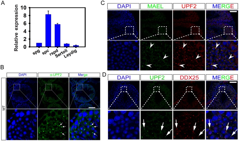
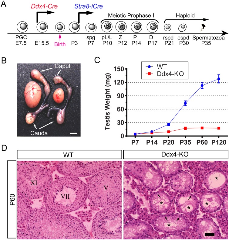
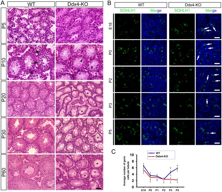
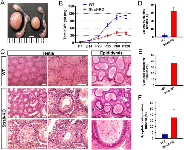
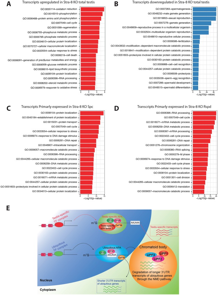
During transcription, most eukaryotic genes generate multiple alternative cleavage and polyadenylation (APA) sites, leading to the production of transcript isoforms with variable lengths in the 3' untranslated region (3'UTR). In contrast to somatic cells, male germ cells, especially pachytene spermatocytes and round spermatids, express a distinct reservoir of mRNAs with shorter 3'UTRs that are essential for spermatogenesis and male fertility. However, the mechanisms underlying the enrichment of shorter 3'UTR transcripts in the developing male germ cells remain unknown. Here, we report that UPF2-mediated nonsense-mediated mRNA decay (NMD) plays an essential role in male germ cells by eliminating ubiquitous genes-derived, longer 3'UTR transcripts, and that this role is independent of its canonical role in degrading "premature termination codon" (PTC)-containing transcripts in somatic cell lineages. This report provides physiological evidence supporting a noncanonical role of the NMD pathway in achieving global 3'UTR shortening in the male germ cells during spermatogenesis.


Conflict of interest statement

The authors have declared that no competing interests exist.

Figures

Fig 1
Fig 1. UPF2 is a novel component of the chromatoid body (CB) and is highly expressed in spermatocytes and round spermatids in murine testes.
(A) qPCR analyses of Upf2 mRNA levels in individual testicular cell types purified from adult murine testes, including spermatogonia (spg), spermatocytes (spc), round spermatids (rspd), Sertoli cells (Sertoli) and Leydig cells (Leydig). Biological triplicates (n = 3) were analyzed and relative Upf2 mRNA levels are shown as means ± SEM. (B) Immunofluorescent localization of UPF2 in WT murine testes. UPF2 is abundantly expressed in the cytoplasm of spermatocytes and round spermatids in WT testes with the highest expression confined to an intensive “dot-like” structure, resembling the chromatoid body (CB, arrows), in round spermatids. Scale bar = 50μm. (C) Double immunofluorescent staining of UPF2 and MAEL, a CB marker, in adult murine testes. Arrowheads indicate CBs in round spermatids. Scale bar = 50μm. (D) Co-localization of UPF2 and DDX25, a CB marker protein. In both stages VIII and I seminiferous tubules, UPF2 mostly overlaps with DDX25 in CBs in the round spermatids (arrows). However, UPF2 is absent in a few DDX25-positive dots (arrowheads), which may represent other types of cytoplasmic granular structures, e.g. the satellite body. Scale bar = 15μm.
Fig 2
Fig 2. UPF2 is essential for prospermatogonial development in murine testes.
(A) A schematic diagram showing the critical time points during male germ cell development. Two Cre deletor lines used express Cre in the male germline starting from E15.5 (Ddx4-Cre) and P4 (Stra8-Cre) as indicated. PGC, primordial germ cell; E, embryonic day; spg, spermatogonium; pL, pre-leptotene spermatocyte; L, leptotene spermatocyte; Z, zygotene spermatocyte; P, pachytene spermatocyte; D, diplotene spermatocyte; rspd, round spermatid; espd, elongating/elongated spermatid. (B) Gross morphology of the testis and the epididymis of WT and Ddx4-KO mice. (C) Testis growth curves of WT and Ddx4-KO mice. Testis weight is presented as means ± SEM (n = 6). (D) Testicular histology of WT and Ddx4-KO mice at P60. Seminiferous tubules of Ddx4-KO mice were devoid of germ cells and suffused with vacuoles (*). Only Sertoli cells (arrows) were present along the basal membrane. Scale bar = 30μm.
Fig 3
Fig 3. Histological analyses of the developing testes in WT and Ddx4-KO mice.
(A) HE staining of paraffin-embedded testicular sections. At postnatal day 5 (P5), the number and morphology of germ cells and Sertoli cells are indistinguishable between WT and Ddx4-KO testes. Upon P10, preleptotene or leptotene spermatocytes (arrows) started to appear in WT testes, but not in Ddx4-KO testes due to germ cell loss and delay in meiotic entry. At P20 and thereafter, numerous vacuoles (*) were present, indicative of massive germ cell depletion in Ddx4-KO testes. Scale bar = 50μm. (B) Loss of prospermatogonia during neonatal testicular development in Ddx4-KO males. Immunostaining of SOHLH1, a marker for prospermatogonia and spermatogonia, in the testes of Ddx4-KO male mice at embryonic day 18 (E18), postnatal day 0 (P0), P2, P3 and P5. Scale bar = 70μm. (C) Quantitative analyses of the average number of germ cells per tubule cross-section in WT and Ddx4-KO testes at E18, P0, P2, P3 and P5. Data are presented as Means ± SD, n = 20. * denotes statistical significance (P<0.05).
Fig 4
Fig 4. UPF2 is required for postnatal germ cell development in mice.
(A) Gross morphology of the testis and the epididymis of WT and Stra8-KO mice. Scale bar = 2mm. (B) Testis growth curve during postnatal development between WT and Stra8-KO mice (n = 6 for each genotype). (C) Histology of WT and Stra8-KO testes and epididymides at P60. While abundant mature spermatozoa were observed in the WT cauda epididymis, only a few degenerating, immature male germ cells were present in the Stra8-KO cauda epididymis. (D-E) Percentage of seminiferous tubules containing vacuoles (D) or multinucleated giant cells (E) in WT and Stra8-KO testes (n = 4 for each genotype). (F) Percentage of tubules containing apoptotic cells based on terminal deoxynucleotidyl transferase dUTP nick end labeling (TUNEL) assay; Scale bar = 50μm.
Fig 5
Fig 5. RNA-Seq analyses reveal no global PTC upregulation, but a drastic increase in the 3’UTR length among upregulated, ubiquitously expressed transcripts in Stra8-KO total testes or purified germ cells.
(A) Heat map showing the clustering of de-regulated transcripts identified through RNA-Seq analyses in Stra8-KO testes. A total of 3,839 transcripts were significantly downregulated, while 1,971 transcripts were significantly upregulated in Stra8-KO total testes (Cutoff: FDR<0.05). (B) PTC analyses of RNA-Seq data demonstrated that among all 23,575 PTC-positive transcripts (17,6% of all transcripts), only 137 were upregulated (~7% of upregulated), suggesting a lack of global PTC upregulation in Stra8-KO testes. All de-regulated genes and transcripts are listed in S1 Table. (C) MA plots of transcript expression showing no global upregulation of PTC-positive transcripts in either Upf2-null spermatocytes (spc) or Upf2-null round spermatids (rspd). Transcripts without annotated start codons (NO ORF), transcripts without PTC (PTC-negative), and transcripts containing PTC (PTC-positive) were analyzed. The blue line indicates the mean log2 fold change along the x-axis. (D) Violin plots showing the length distribution of full-length transcript, ORF, 5’UTRs and 3’UTR among up- (1,559), down- (2,654) and non-regulated (85,910) transcripts in Stra8-KO testes (Cutoff: FDR < 0.05). Only transcripts derived from genes expressing multiple transcripts were analyzed. The three dots in each violin plot indicate the 25th, 50th (median) and 75th percentile of the visualized data. (E) Statistics for the pairwise comparisons of the length distributions shown in D. The most significant length increases were observed in 3’UTRs of the up-regulated transcripts in Stra8-KO testes (p-value = 1.05e-149, between up- and down- groups). (F) The median 3’UTR length of the up-, down- and non-regulated transcripts analyzed in D.
Fig 6
Fig 6. Longer 3’UTR, not PTC-containing transcripts, were accumulated in Upf2-null spermatocytes and round spermatids.
(A) Bar plot showing the number of transcripts primarily expressed in WT, Stra8-KO or both genotypes, in spermatocytes (spc) and round spermatids (rspd). Cutoff: 1 normFPKM (see Methods and Materials). Classification of genes primarily expressed in WT, KO or both is given in S1 Table. (B) Violin plots showing the length distribution of the full-length transcripts, ORFs, 5’UTRs and 3’UTRs from the transcripts primarily expressed in WT, Stra8-KO or both genotypes. Rows indicate spermatocytes (spc) and round spermatids (rspd). The three dots in each violin plot indicate the 25th, 50th (median) and 75th percentile of the visualized data. Only transcripts derived from genes expressing multiple transcripts were analyzed. (C) A summary of statistics for the pairwise comparisons of the length distributions shown in B. (D) Distribution of the percentage by which each individual transcript contribute to their respective parent gene as measured in WT (red) and Stra8-KO (blue) in spermatocytes (left column) and round spermatids (right column). The analysis was performed for transcripts primarily expressed in WT, Stra8-KO or both genotypes (rows). Only transcripts derived from genes expressing multiple transcripts were analyzed. (E) A summary of statistics for the pairwise comparison of the length distributions shown in D. (F) Illustration of the strategy used for semi-quantitative PCR-based validation, with one pair from the protein-coding region (primer set 1) and the other from the 3’UTRs (primer set 2). (G) Semi-quantitative PCR-based validation of four randomly chosen transcripts with longer 3’UTRs (Pank3, Gpx3, Map4k4 and Klf6). Note the alternative transcripts with longer 3’UTRs were readily detected in Stra8-KO samples but not in WT samples.
Fig 7
Fig 7. UPF2 selectively eliminates longer 3’UTR transcripts derived from ubiquitously expressed genes in spermatocytes and round spermatids.
(A-B) Gene ontology (GO) analyses of differentially expressed genes (FDR < 0.05) for up- and down-regulated genes respectively (P<0.01; top 18 GO terms are shown). (C-D) Gene ontology (GO) analyses of genes with transcripts selectively upregulated upon Upf2 inactivation in spermatocytes (Spc) and round spermatids (Rspd) respectively (P<0.01; top 18 GO terms are shown). (E) A working model for UPF2-mediated 3’UTR length control in male germ cells. While the testis-specific transcription factor (TF) complex containing the yet-to-be-identified testis-specific APA factors produces testis-specific gene transcripts with shorter 3’UTRs, the ubiquitous TF complex cooperates with the ubiquitous APA complex to generate both shorter and longer 3’UTR transcripts from ubiquitously expressed genes in male germ cells. The transcripts with longer 3’UTRs are then selectively degraded by the UPF2-directed NMD in the chromatoid body, leading to enrichment of shorter 3’UTR transcripts in haploid male germ cells.

Comment in

Similar articles

Cited by

References

    1. Clermont Y. Kinetics of spermatogenesis in mammals: seminiferous epithelium cycle and spermatogonial renewal. Physiological reviews. 1972;52(1):198–236. Epub 1972/01/01. . - PubMed
    1. Braun RE. Post-transcriptional control of gene expression during spermatogenesis. Seminars in cell & developmental biology. 1998;9(4):483–9. Epub 1998/11/14. 10.1006/scdb.1998.0226 . - DOI - PubMed
    1. Eddy EM. Male germ cell gene expression. Recent progress in hormone research. 2002;57:103–28. Epub 2002/05/23. . - PubMed
    1. Fajardo MA, Haugen HS, Clegg CH, Braun RE. Separate elements in the 3' untranslated region of the mouse protamine 1 mRNA regulate translational repression and activation during murine spermatogenesis. Developmental biology. 1997;191(1):42–52. Epub 1997/11/14. 10.1006/dbio.1997.8705 . - DOI - PubMed
    1. Kleene KC. Patterns, mechanisms, and functions of translation regulation in mammalian spermatogenic cells. Cytogenetic and genome research. 2003;103(3–4):217–24. Epub 2004/03/31. 76807. . - PubMed

MeSH terms