Review
. 2016 Sep;31(5):253-72.
doi: 10.1097/RTI.0000000000000217.

Cardiothoracic Applications of 3-dimensional Printing

Affiliations
Review

Cardiothoracic Applications of 3-dimensional Printing

Andreas A Giannopoulos et al. J Thorac Imaging. 2016 Sep.

Abstract

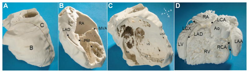
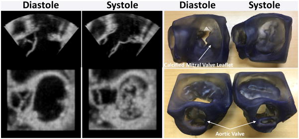
Medical 3-dimensional (3D) printing is emerging as a clinically relevant imaging tool in directing preoperative and intraoperative planning in many surgical specialties and will therefore likely lead to interdisciplinary collaboration between engineers, radiologists, and surgeons. Data from standard imaging modalities such as computed tomography, magnetic resonance imaging, echocardiography, and rotational angiography can be used to fabricate life-sized models of human anatomy and pathology, as well as patient-specific implants and surgical guides. Cardiovascular 3D-printed models can improve diagnosis and allow for advanced preoperative planning. The majority of applications reported involve congenital heart diseases and valvular and great vessels pathologies. Printed models are suitable for planning both surgical and minimally invasive procedures. Added value has been reported toward improving outcomes, minimizing perioperative risk, and developing new procedures such as transcatheter mitral valve replacements. Similarly, thoracic surgeons are using 3D printing to assess invasion of vital structures by tumors and to assist in diagnosis and treatment of upper and lower airway diseases. Anatomic models enable surgeons to assimilate information more quickly than image review, choose the optimal surgical approach, and achieve surgery in a shorter time. Patient-specific 3D-printed implants are beginning to appear and may have significant impact on cosmetic and life-saving procedures in the future. In summary, cardiothoracic 3D printing is rapidly evolving and may be a potential game-changer for surgeons. The imager who is equipped with the tools to apply this new imaging science to cardiothoracic care is thus ideally positioned to innovate in this new emerging imaging modality.


Conflict of interest statement

Dr. Mitsouras receives research support from Vital Images, A Toshiba Medical Systems Group Company

Figures

Figure 1
Figure 1. Generation of a 3D-printable STL model from a volumetric medical image dataset
The aorta and aortic arch vessels are first segmented from a contrast enhanced CT (A). The segmented image voxels identify the region of space occupied by blood and conversely this region of space is entirely filled by the individually-segmented voxels (B). If one were to cut through this region, it would simply expose the inner voxels that have been segmented (C). An STL model that can be 3D-printed is instead a surface composed of small triangles that encloses the segmented voxels (D; shown in red, with individual triangle outlines shown in inset). Cutting this surface merely exposes the inner side of the triangles (E; shown in green, with individual triangle outlines shown in inset).
Figure 2
Figure 2. Addition of non-anatomic features to STL models of the clavicle and ribs segmented from a CT for inclusion in a superior sulcus tumor model
The model does not include the sternum to simplify interpretation by the surgical team. To maintain the relative position of the clavicle to the rib, two cylindrical connectors are added between the rib and clavicle STLs using CAD tools (red arrows). After printing, the two structures will stay united in one piece.
Figure 3
Figure 3. Complete sequence of STL generation and CAD manipulations to produce a model for planning of an endovascular procedure in an aorta with mural thrombi from contrast-enhanced CTA
CTA sagittal plane illustrates the contrast-enhanced lumen and thrombi (A). Segmentation of the blood pool and thrombus (B; lumen in red, thrombus in green) yields the corresponding STLs (C,E). The STL surfaces can be viewed superposed on the original CTA (D,F). These STL files can be printed but would not allow appreciating the extent of thrombus volume and lumen loss that are important for planning the procedure. An ideal 3D-printed model would show a hollow lumen with the thrombi extruding into it. This is achieved by first using “wrapping” and “smoothing” CAD manipulations on the blood pool STL to produce the hypothetical ideal, smooth lumen (G,H; hypothetical lumen STL superposed on CT in bold red, and initial blood pool STL in lighter red). From this ideal lumen, a “hollow” procedure is applied to extrude a virtual vessel wall outwards from the lumen (I,J; virtual wall in turquoise). The mural thrombi now extend from the wall into the hollow lumen (K,L) as intended. A final CAD manipulation is used to create a cutout window in the vessel wall (M) to allow inspection of the lumen (N). The cutout portion is further augmented with snap-fit connectors using CAD tools to allow attachment to the main model (O,P). Same case as shown in Figure 1; the final 3D-printed model is shown in Figure 11.
Figure 4
Figure 4. Congenital heart disease models for surgical simulation
Panel A depicts 3D printed model of a case of criss-cross heart with double outlet right ventricle. Upper row, shows the printout of the blood pool. Lower row shows a hollow model of the same case with a fictitious wall around the contrast-enhanced blood pool representing an approximation of the vessels and myocardium wall. Panel B shows a 3D printed model of a hypoplastic left ventricle heart syndrome during mockup operation for training and simulation of the Norwood operation. Figure courtesy of Prof. Shi-Joon Yoo from The Hospital for Sick Children, University of Toronto, Toronto, Canada.
Figure 5
Figure 5. MRI-derived physical model of complex CHD
The 3D printed model of the myocardium plus vessel walls (Panel A) consists of two pieces (Panels B and C). A single cut divides the right atrium (RA) and the left ventricle (LV), allowing the view on the mitral valve annulus (MVA), two papillary muscles (PM), the left anterior descending coronary artery (LAD), ventricular septal defect (VSD), ventricular septum (VS), atrial septal defect (ASD), and the aorta (Ao). Panel D shows a 3D-printed model of the intracardiac volumes (blood pool) from the left anterior view. There is atrioventricular discordance with connections of the right atrium (RA) to the morphologically left ventricle (LV) on the right side. The left atrium (LA) with an enlarged left atrial appendage (LAA) is connected with the morphologically right ventricle (RV) on the left side. A dilated aorta (Ao) rises from the right ventricle. The left coronary artery (LCA) and right coronary artery (RCA) rise from a common origin of the coronary arteries (CO). (LAD = left anterior descending coronary artery; LCX = left circumflex; RV = right ventricle, A= anterior; L = left; P = posterior; R = right.) Reprinted with permission from reference [5].
Figure 6
Figure 6. 3D printing of complementary models for vascular pathologies
Representative example of a renal aneurysm contrast-enhanced CTA (A) used to produce 3D-printed models of the blood pool (B) and lumen for the left renal artery aneurysm (C). After examining the models, the surgeons opted for open repair of the aneurysm and patch angioplasty.
Figure 7
Figure 7. Models for planning and simulation of stent deployment for Mustard baffle revision in 45 year old male with history of complete transposition of great vessels
Panel A: Delayed venous phase CT demonstrating a large defect between the IVC and the pulmonary venous pathways at the rightward aspect of the baffle, a smaller defect between the SVC and the pulmonary venous pathways, and an intermediate-sized defect between the baffle and the right atrial appendage (red arrows). Panel B: 3D-printed model of the baffle designed as a fictitious wall around the blood pool (printed in gray) and including the ventricles (printed in white) for spatial orientation in this difficult case. Panel C: Removable ventricles and cut-out window of the wall of the pulmonary venous pathway/right atrium demonstrate the superior small and inferior large baffle defect (red arrows) and cut-out window of the right atrial wall demonstrates the third baffle defect communicating with the right atrial appendage. Panel D: A segment of the baffle was also printed in flexible material and used to simulate stent graft deployment to ensure an adequate proximal sealing zone. PV: Pulmonary vein, SVC: Superior vena cava, IVC: Inferior vena cava, RA: Right atrium, RV: Right ventricle, LV: left ventricle, RAA: Right atrial appendage.
Figure 8
Figure 8. 3D printing of stenotic aortic valve for functional modeling
Top panel: clear model designed from patient CT examination indicates the location of a calcified aortic valve leaflet. Middle panel: Similar 2D echocardiographic image quality between patient and model with clear depiction of aortic root geometry and aortic valve leaflet calcification (red arrow). Bottom panel: Continuous wave Doppler imaging (matched to stroke volume and pressure gradient) demonstrates similar peak velocity, ejection time, and overall Doppler signal quality for patient and 3D printed model. Reprinted with permission from reference [55].
Figure 9
Figure 9. 3D printing of mitral and aortic valves from 3D TEE
Valves 3D printed in end-diastole and end-systole from lightly smoothed 3D TEE images to reduce speckles. Although the static models cannot assist in appreciating valve function, they clearly demonstrate the calcified middle posterior mitral valve leaflet impeding flow into the left ventricle. The models can thus be used to appreciate geometry and perform accurate measurements.
Figure 10
Figure 10. Three-dimensional echocardiography, virtual, and physical models of a patient with severe mitral regurgitation and a flail P2 leaflet segment
Long-axis view of the left ventricle (LV) and atrium during systole (A). The relative positions of the anterior (AL) and posterior (PL) mitral valve leaflets are indicated. Virtual (B) and printed model (C) viewed from the atrium. Virtual (D) and printed model (E) as viewed in profile from anterior to posterior commissure. Reprinted with permission from reference [61].
Figure 11
Figure 11. Model of aortic wall with mobile mural thrombi generated from CTA
The contrast-enhanced aorta with the mural thrombi seen as filling defects (A, green arrows) and high-density calcifications (A, white arrowheads). 3D volume rendering of the CTA conveys the locations of the calcifications and thrombi (B). A printed model of the aortic wall seen from the exterior (C) includes a cutout window that can be removed to inspect the aortic lumen (D). Inspection of the model from the cutout window (E,F) and viewed upwards from the descending aorta (G) allows appreciation of the location and size of the calcifications and thrombi (E-G, white arrowheads and green arrows, respectively) including with relation to the aortic arch vessels for planning of the percutaneous intervention.
Figure 12
Figure 12. Models of the tracheobronchial tree of a 67 year old man with known relapsing polychondritis
Top row: anterior and posterior views of the inspiratory phase; bottom row: anterior and posterior views in expiration, evidently showing the collapsed airways. Reprinted with permission from reference [8].
Figure 13
Figure 13. Airway model for planning of single-lung ventilation in a small child
The model allowed experimentation with various sizes of tracheal tube. In this example a size 3.5 Microcuff® tube was used, with cuff placed in the trachea (A) and with its tip at the carina, as well as a size 4 Microcuff® tube (B), with cuff placed in the left main bronchus. Reprinted with permission from reference [71].
Figure 14
Figure 14. 3D-Printed custom-made titanium prosthesis for sternocostal reconstruction
CT scan showing involvement of chest wall structures (A). Final 3D printed implant design (B). A rigid template to allow precise setting of resection margins 3D-printed from biocompatible material is placed in the operative field (C) and final placement of the prosthesis with a Dualmesh® patch fixed to its rear side (D). Reprinted with permission from reference [16].
Figure 15
Figure 15. 3D printing of esophageal pathology
Patient with multiple esophageal diverticula imaged using a novel positive oral contrast and air technique to delineate the esophagus (A). The esophagus, stomach, diaphragmatic crus, aorta, and spine are individually segmented (B; each structure shown in different color). STL models of each segmented structure (C) are then 3D-printed in a multi-material, multi-colored model was printed (D). Reprinted with permission from reference [76].
Figure 16
Figure 16. Model of a superior sulcus tumor
Contrast enhanced CT demonstrating the segmentation of the aorta in a coronal plane (A), and the pulmonary vasculature (B) and apical lung mass (C) in a sagittal plane. The complete STL (D) and 3D-printed models (E) include the mass, left 1st-4th ribs and upper thoracic vertebrae, thoracic aorta and great vessels, central and left upper lobe pulmonary vasculature, left subclavian and bilateral brachiocephalic veins, and the superior vena cava.
Figure 17
Figure 17. Model of middle mediastinal mass
Model includes the aorta, pulmonary artery, superior vena cava, and tracheobronchial tree, demonstrating a lobulated middle mediastinal mass extending from right paratracheal region to the aortopulmonary window and insinuating between the aorta and the main pulmonary artery.

References

    1. Markl M, Schumacher R, Kuffer J, et al. Rapid vessel prototyping: vascular modeling using 3t magnetic resonance angiography and rapid prototyping technology. MAGMA. 2005;18(6):288–92. - PubMed
    1. McGurk M, Amis AA, Potamianos P, et al. Rapid prototyping techniques for anatomical modelling in medicine. Annals of The Royal College of Surgeons of England. 1997;79(3):169–174. - PMC - PubMed
    1. Noecker AM, Chen JF, Zhou Q, et al. Development of patient-specific three-dimensional pediatric cardiac models. ASAIO J. 2006;52(3):349–53. - PubMed
    1. Schievano S, Sala G, Migliavacca F, et al. Use of rapid prototyping models in the planning of percutaneous pulmonary valved stent implantation. Proceedings of the Institution of Mechanical Engineers, Part H: Journal of Engineering in Medicine. 2007;221(4):407–416. - PubMed
    1. Mottl-Link S, Hubler M, Kuhne T, et al. Physical models aiding in complex congenital heart surgery. Ann Thorac Surg. 2008;86(1):273–7. - PubMed