Observational Study
. 2016 May;95(18):e3466.
doi: 10.1097/MD.0000000000003466.

The Kinetics of Circulating Monocyte Subsets and Monocyte-Platelet Aggregates in the Acute Phase of ST-Elevation Myocardial Infarction: Associations with 2-Year Cardiovascular Events

Affiliations
Observational Study

The Kinetics of Circulating Monocyte Subsets and Monocyte-Platelet Aggregates in the Acute Phase of ST-Elevation Myocardial Infarction: Associations with 2-Year Cardiovascular Events

Xin Zhou et al. Medicine (Baltimore). 2016 May.

Abstract

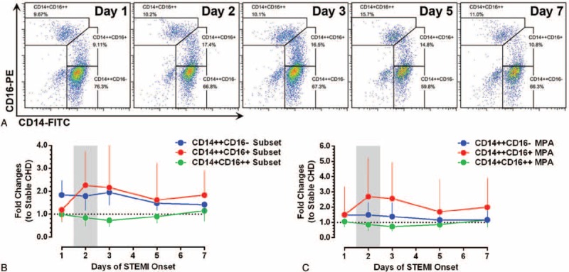
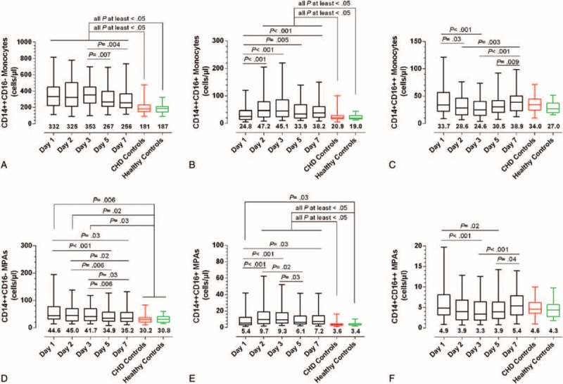
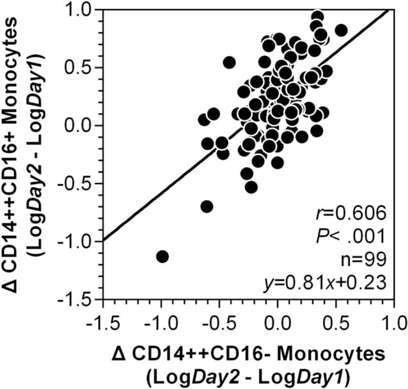
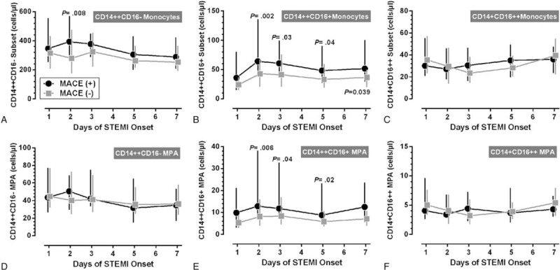
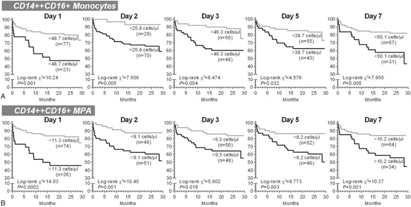
In experimental myocardial infarction (MI), a rise in cell counts of circulating monocyte subsets contributes to impaired myocardial healing and to atherosclerotic plaque destabilization. In humans, the prognostic role of monocyte subsets in patients suffering ST-elevation MI (STEMI) is still unclear. In the present study, we aimed to determine the kinetics of the 3 monocyte subsets (classical CD14++CD16-, intermediate CD14++CD16+, and nonclassical CD14+CD16++ monocytes), as well as the subset-specific monocyte-platelet aggregates (MPA), in acute STEMI followed by primary percutaneous coronary intervention (PCI), and their relationships with cardiovascular outcomes during a 2-year follow-up.Monocyte subsets and MPA were measured in 100 STEMI patients receiving primary PCI on days 1, 2, 3, 5, and 7 of symptom onset, which were compared with 60 stable coronary heart disease patients and 35 healthy volunteers. From day 1 to day 7, significant increases in the counts of CD14++CD16+ monocytes and CD14++CD16+ MPA were observed, with peak levels on day 2. During a median follow-up of 2.0 years, 28 first cardiovascular events (defined as cardiovascular death, nonfatal ischemic stroke, recurrent MI, need for emergency or repeat revascularization, and rehospitalization for heart failure) were recorded. After adjustment for confounders, CD14++CD16+ monocytosis (day 1 [HR: 3.428; 95% CI: 1.597-7.358; P = 0.002], day 2 [HR: 4.835; 95% CI: 1.106-21.13; P = 0.04], day 3 [HR: 2.734; 95% CI: 1.138-6.564; P = 0.02], and day 7 [HR: 2.647; 95% CI: 1.196-5.861; P = 0.02]), as well as increased levels of CD14++CD16+ MPA measured on all time points (days 1, 2, 3, 5, and 7), had predictive values for adverse cardiovascular events.In conclusion, our data show the expansion of the CD14++CD16+ monocyte subset during acute phase of STEMI has predictive values for 2-year adverse cardiovascular outcomes in patients treated with primary PCI. Future studies will be warranted to elucidate whether CD14++CD16+ monocytes may become a target cell population for new therapeutic strategies after STEMI.


Conflict of interest statement

The authors have no conflicts of interest to disclose.

Figures

FIGURE 1
FIGURE 1
The dynamics of monocyte subsets and subset-specific MPA after STEMI. (A) Illustrate the dynamic changes of monocyte subset in a single patient (FCM analysis; representative example). (B, C) The lower panel shows the temporal profiles of monocyte subsets and subset-specific MPA in STEMI patients (fold changes to stable coronary heart disease patients). Data are presented as medians and interquartile ranges. MPA = monocyte–platelet aggregates, STEMI = ST-elevation myocardial infarction.
FIGURE 2
FIGURE 2
The monocyte subsets and subset-specific MPA in STEMI and coronary heart disease patients, and in healthy controls. (A–C) Changes in CD14++CD16−, CD14++CD16+, and CD14+CD16++ monocytes, respectively. (D–F) Changes in CD14++CD16−, CD14++CD16+, and CD14+CD16++ MPA, respectively. The data are presented by box and whisker plots: the boxes extend from the 25th to the 75th percentile, with a line at the median. The whiskers extend above and below the box to show the 5th to 95th percentiles of values. The number below each box and whisker plot shows the median value. MPA = monocyte–platelet aggregates, STEMI = ST-elevation myocardial infarction.
FIGURE 3
FIGURE 3
Correlation between ΔCD14++CD16− monocytes and ΔCD14++CD16+ monocytes after ST-elevation myocardial infarction. The Δ value was defined as the value measured on day 2 minus the value measured on day 1 of ST-elevation myocardial infarction onset. The data are presented after log transformation. Correlation coefficients are reported as Pearson linear correlations.
FIGURE 4
FIGURE 4
Monocyte subsets and subset-specific MPA between MACE(+) and MACE(−) STEMI patients. (A–C) Changes in CD14++CD16−, CD14++CD16+, and CD14+CD16++ monocytes in MACE(+) and MACE(−) patients, respectively. (D–F) Changes in CD14++CD16−, CD14++CD16+, and CD14+CD16++ MPA in MACE(+) and MACE(−) patients, respectively. Data are presented as medians and interquartile ranges. The P values indicate the results of statistical comparisons between MACE(+) and MACE(−) patients at the same time point. MACE = major adverse cardiovascular events, MPA = monocyte–platelet aggregates, STEMI = ST-elevation myocardial infarction.
FIGURE 5
FIGURE 5
The univariate Kaplan–Meier survival analyses for 2-year major adverse cardiovascular events. The patients were stratified according the optimal cut-off values derived from receiver operator characteristic curve analyses. MPA = monocyte-platelet aggregates.

References

    1. Frangogiannis NG. The inflammatory response in myocardial injury, repair, and remodelling. Nat Rev Cardiol 2014; 11:255–265. - PMC - PubMed
    1. Seropian IM, Toldo S, Van Tassell BW, et al. Anti-inflammatory strategies for ventricular remodeling following ST-segment elevation acute myocardial infarction. J Am Coll Cardiol 2014; 63:1593–1603. - PubMed
    1. Zhou X, Yun JL, Han ZQ, et al. Postinfarction healing dynamics in the mechanically unloaded rat left ventricle. Am J Physiol Heart Circ Physiol 2011; 300:H1863–H1874. - PMC - PubMed
    1. Ruparelia N, Godec J, Lee R, et al. Acute myocardial infarction activates distinct inflammation and proliferation pathways in circulating monocytes, prior to recruitment, and identified through conserved transcriptional responses in mice and humans. Eur Heart J 2015; 36:1923–1934. - PMC - PubMed
    1. Swirski FK, Nahrendorf M. Leukocyte behavior in atherosclerosis, myocardial infarction, and heart failure. Science 2013; 339:161–166. - PMC - PubMed

Publication types

MeSH terms