. 2016 May 5;11(5):e0154412.
doi: 10.1371/journal.pone.0154412. eCollection 2016.

A Mutation in LTBP2 Causes Congenital Glaucoma in Domestic Cats (Felis catus)

Affiliations

A Mutation in LTBP2 Causes Congenital Glaucoma in Domestic Cats (Felis catus)

Markus H Kuehn et al. PLoS One. .

Erratum in

Abstract

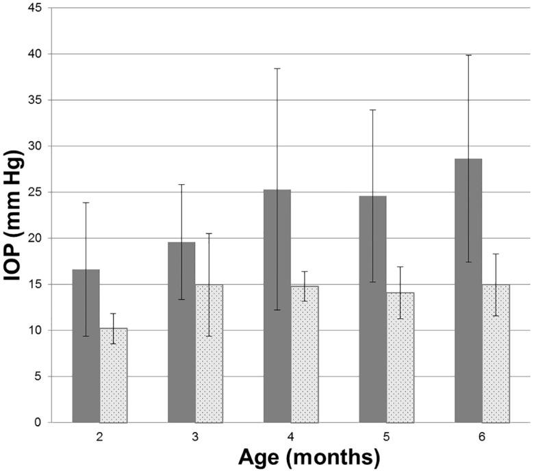
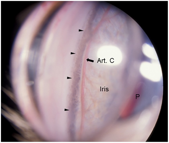
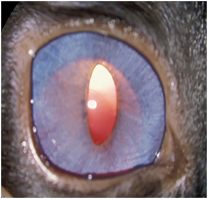
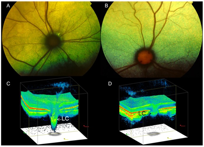
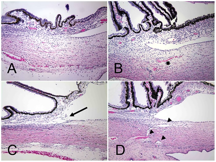
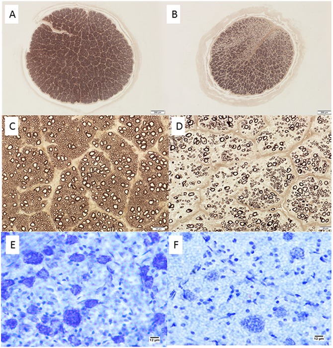
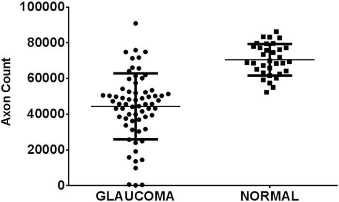
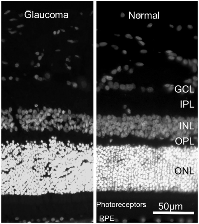
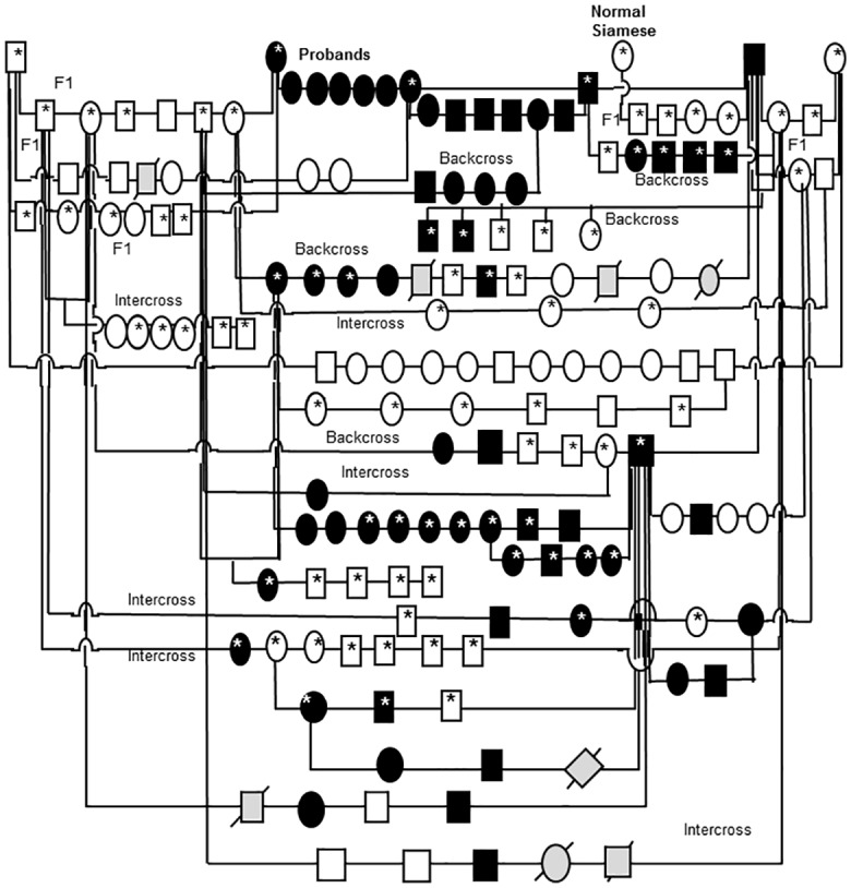
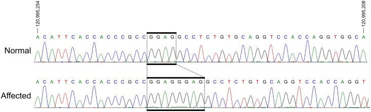
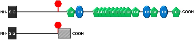
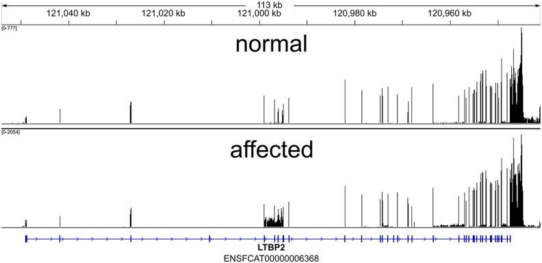
The glaucomas are a group of diseases characterized by optic nerve damage that together represent a leading cause of blindness in the human population and in domestic animals. Here we report a mutation in LTBP2 that causes primary congenital glaucoma (PCG) in domestic cats. We identified a spontaneous form of PCG in cats and established a breeding colony segregating for PCG consistent with fully penetrant, autosomal recessive inheritance of the trait. Elevated intraocular pressure, globe enlargement and elongated ciliary processes were consistently observed in all affected cats by 8 weeks of age. Varying degrees of optic nerve damage resulted by 6 months of age. Although subtle lens zonular instability was a common feature in this cohort, pronounced ectopia lentis was identified in less than 10% of cats examined. Thus, glaucoma in this pedigree is attributed to histologically confirmed arrest in the early post-natal development of the aqueous humor outflow pathways in the anterior segment of the eyes of affected animals. Using a candidate gene approach, significant linkage was established on cat chromosome B3 (LOD 18.38, θ = 0.00) using tightly linked short tandem repeat (STR) loci to the candidate gene, LTBP2. A 4 base-pair insertion was identified in exon 8 of LTBP2 in affected individuals that generates a frame shift that completely alters the downstream open reading frame and eliminates functional domains. Thus, we describe the first spontaneous and highly penetrant non-rodent model of PCG identifying a valuable animal model for primary glaucoma that closely resembles the human disease, providing valuable insights into mechanisms underlying the disease and a valuable animal model for testing therapies.


Conflict of interest statement

Competing Interests: The authors have declared that no competing interests exist.

Figures

Fig 1
Fig 1. Intraocular pressure in glaucomatous and normal cats.
Intraocular pressure measured by rebound tonometry (TonoVet, ICare oy, Finland) was significantly greater in PCG (solid bars) than in normal cats (patterned bars) at all ages from 2 months of age (ANOVA with Tukey-Kramer multiple comparisons post-test, p<0.05). Error bars represent standard deviation. Numbers (n) of subjects in each age group are indicated on each column.
Fig 2
Fig 2. Gonioscopic Features of Feline Primary Congenital Glaucoma.
Goniophotograph illustrates an open, but slightly narrowed, iridocorneal angle (opening of ciliary cleft; indicated by arrow heads) and mild dysplasia of the pectinate ligament visualized by gonioscopy in a 7 month-old Siamese cat with primary congenital glaucoma. No ectopia lentis was evident in this subject. (I = Iris, P = pupil, Art. C = major arterial circle of iris).
Fig 3
Fig 3. Clinical Appearance of Feline Primary Congenital Glaucoma.
Iris hypoplasia and ectopia lentis in feline PCG. The outline of the posteriorly subluxated lens can be readily appreciated through thin, translucent iris tissue in this affected Siamese cat.
Fig 4
Fig 4. Optic Nerve Head Cupping in Feline Primary Congenital Glaucoma.
Fundus photographs illustrate the cupped and degenerate optic nerve head of [A] a cat with advanced primary congenital glaucoma and compared with a normal age-matched cat [B]. An optic nerve cube scan obtained by spectral domain optical coherence tomography (OCT;Cirrus, Carl Zeiss Meditec Inc., Dublin, CA) acquired in this affected cat confirms dramatic posterior displacement of the lamina cribrosa (LC, arrowed)[C] compared to the normal control[D]. (Modified, with permission, from McLellan GJ, Rasmussen CA. Optical coherence tomography for the evaluation of retinal and optic nerve morphology in animal subjects: practical considerations. Veterinary Ophthalmology. Sep 2012;15 Suppl 2:13–28.).
Fig 5
Fig 5. Arrested development of Aqueous Humor Outflow Pathways in Feline Primary Congenital Glaucoma.
Photomicrographs of the iridocorneal angle in one day old (A and B) and 18 day old (C and D) cats. Affected cats (A and C) from the pedigree appear to develop fewer scleral vessels (asterisk) than normal and F1 out-crossed animals (B and D). Affected animals also fail to develop the normal vessels of the angular aqueous plexus (analogous to the canal of Schlemm in humans), collector channels and intrascleral venous plexus (arrowheads).
Fig 6
Fig 6. Retinal Ganglion Cell Loss in Cats With Primary Congenital Glaucoma.
Photomicrographs showing representative optic nerve sections [A-D], stained with p-phenylenediamine to highlight axonal myelin sheaths, from 2 young adult cats. [A,C] normal cat (estimated 83,398 optic nerve axons); [B,D] typical PCG-affected cat with moderate degree of axon loss (estimated 30,365 optic nerve axons). Mid-peripheral retina in cresyl violet stained retinal whole mounts from these same two cats show relative loss of Nissl-stained retinal ganglion cell (RGC) somas of >12μm diameter in the ganglion cell layer of the retina in PCG (total RGC soma count in whole retina of 43,891)[E] relative to a normal cat [F] (estimated RGC soma count in whole retina of 123,833). Bar markers = 200μm (top row); 20μm (middle row), and 12μm (bottom row).
Fig 7
Fig 7. Optic Neuropathy in Feline Primary Congenital Glaucoma.
Mean axon count in PCG cats (66 eyes of 38 cats aged 4 months to 8 years) is significantly lower (44,438 ± 2,272 [SEM]) than in normal cats (mean = 70,510 ± 1,535 in 33 eyes of 20 normal cats; p<0.0001, unpaired t-test). Lines represent mean and error bars represent SD.
Fig 8
Fig 8. Preservation of Normal Outer Retinal Structure in Feline Primary Congenital Glaucoma.
Representative fluorescence photomicrographs of DAPI stained 18 month old glaucomatous [A] and normal 6 month old [B] feline retinas. No significant difference in morphology of outer retinal layers is identified. (GCL = ganglion cell layer; IPL = inner plexiform layer; INL = inner nuclear layer; OPL = outer plexiform layer; ONL = outer nuclear layer; RPE = retinal pigment epithelium).
Fig 9
Fig 9. Feline congenital glaucoma pedigree.
Additional pairings between affected and affected animals consistently produced all affected offspring but are not depicted in this informative pedigree. Females are depicted by circles and males by squares, diamonds indicate gender not recorded. Black shapes indicate that PCG phenotype confirmed by clinical examination conducted by a board certified veterinary ophthalmologist. Unfilled shapes indicate normal phenotype confirmed on clinical examination. Light gray shading with strike-through indicates that phenotype was not confirmed. Asterisks indicate those animals from which DNA samples were obtained and used in linkage analysis and/ or gene sequencing.
Fig 10
Fig 10. Sequences of Exon 8 of LTBP2 in Normal Cat and in Primary Congenital Glaucoma.
Sanger sequencing results of exon 8 of LTBP2 in a normal (top) and a PCG-affected (bottom) individual. Analysis reveals a 4bp insertion in all affected individuals (boxed). Coordinates are for chromosome B3 from the felCat5 genome build.
Fig 11
Fig 11. Predicted Effects of Mutation on Feline LTBP2.
Schematic representation of the feline LTBP2 protein in normal (top) and affected cats (bottom). The sequence contains an amino terminal signal sequence (dark rectangle), 13 EGF-like calcium binding domains (green) and 3 TGF binding domains (blue). The insertion site causing a frameshift mutation in affected animals is marked by a vertical bar and the altered sequence caused by the frameshift is indicated by a hatched box.
Fig 12
Fig 12. Coverage across Feline LTBP2 exons by RNA-Seq.
RNA-Seq read coverage across the LTBP2 exons within the cat genome. Coordinates are shown in genomic orientation. The numeric range in the top left indicates the minimum and maximum number of reads (FPKM-Fragments Per Kilobase of exon per Million) per base for the sample.

References

    1. Tham YC, Li X, Wong TY, Quigley HA, Aung T, Cheng CY. Global prevalence of glaucoma and projections of glaucoma burden through 2040: A systematic review and meta-analysis. Ophthalmology. 2014. June 26 . - PubMed
    1. Leske MC, Heijl A, Hussein M, Bengtsson B, Hyman L, Komaroff E. Factors for glaucoma progression and the effect of treatment: The early manifest glaucoma trial. Archives of Ophthalmology. 2003. January;121(1):48–56. . - PubMed
    1. Kong L, Fry M, Al-Samarraie M, Gilbert C, Steinkuller PG. An update on progress and the changing epidemiology of causes of childhood blindness worldwide. Journal of American Association for Pediatric Ophthalmology and Strabismus. 2012. 12//;16(6):501–507. 10.1016/j.jaapos.2012.09.004 - DOI - PubMed
    1. Lim S-H, Tran-Viet K-N, Yanovitch TL, Freedman SF, Klemm T, Call W, et al. CYP1B1, MYOC, and LTBP2 mutations in primary congenital glaucoma patients in the united states. American Journal of Ophthalmology. 2013. 3//;155(3):508–517.e505. 10.1016/j.ajo.2012.09.012 - DOI - PMC - PubMed
    1. Alodhayb S, Morales J, Edward DP, Abu-Amero KK. Update on pediatric glaucoma. Seminars in Ophthalmology. 2013. May;28(3):131–143. 10.3109/08820538.2013.771196 - DOI - PubMed

Publication types

Substances