. 2016 Apr 26;15(4):774-786.
doi: 10.1016/j.celrep.2016.03.061. Epub 2016 Apr 14.

A Unique Class of Neural Progenitors in the Drosophila Optic Lobe Generates Both Migrating Neurons and Glia

Affiliations

A Unique Class of Neural Progenitors in the Drosophila Optic Lobe Generates Both Migrating Neurons and Glia

Zhenqing Chen et al. Cell Rep. .

Abstract

How neuronal and glial fates are specified from neural precursor cells is an important question for developmental neurobiologists. We address this question in the Drosophila optic lobe, composed of the lamina, medulla, and lobula complex. We show that two gliogenic regions posterior to the prospective lamina also produce lamina wide-field (Lawf) neurons, which share common progenitors with lamina glia. These progenitors express neither canonical neuroblast nor lamina precursor cell markers. They bifurcate into two sub-lineages in response to Notch signaling, generating lamina glia or Lawf neurons, respectively. The newly born glia and Lawfs then migrate tangentially over substantial distances to reach their target tissue. Thus, Lawf neurogenesis, which includes a common origin with glia, as well as neuronal migration, resembles several aspects of vertebrate neurogenesis.


Figures

Figure 1
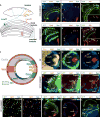
Figure 1. Lamina Wide-Field Neurons Express Specific Transcription Factors
(A) Lamina wide-field neurons have cell bodies in the medulla cortex and arborizations in both the lamina and medulla neuropils. Lawf1 cells (blue) project to layers M1 and M3 of the medulla, whereas Lawf2 cells (magenta) project to M1 and M9. The locations of adult epithelial glia (eg) and marginal glia (mg) are also shown. Adapted from Tuthill et al. (2013) and Edwards et al. (2012). (B and C) 1118-Gal4 drives expression in Lawf1 neurons in adults. Lawf1s express Eya (blue in B) and Hth (red in C) but not Lim1 (red in B). (D–E) 11D03-Gal4 drives expression in Lawf2s in adults (green). Lawf2 neurons express Lim1 (red in D) and Hth (red in E). While most Lawf2 neurons express Eya (arrows, magenta cells in D), some do not (arrowheads in D and D′). (F) Schematic drawing of larval optic lobe and Lawf positions (sagittal cross section). (G) In larval brains, Lawf1 neurons express Hth (green) and Eya (blue); Lawf2 neurons express Lim1, Hth, and Eya. (H–K′) 17C11-Gal4 drives expression in both Lawf1s and Lawf2s. (H) In a later larval brain, all Lawf neurons are Eya+. (I) At the prepupal stage, the two Lawf populations move toward each other (curved arrows). (J) One day into pupation, Lim1+ Lawf2 and Lim1 Lawf1 populations migrate to populate the entire medulla cortex and intermingle with each other. Eya is lost in some Lawf2 neurons (arrows). (K) Two days into pupation, Lawf1 and Lawf2 populations form their arborization patterns in the lamina and medulla neuropils. 17C11-Gal4 expression is reduced in Lawf2s. Blue arrow points to a Lawf1 cell body. Blue arrowhead points to Lawf1 projections in the M3 layer. Magenta arrow points to a Lawf2 cell body. Magenta arrowhead points to Lawf2 projections in the M3 layer. (L) In the adult, 17C11-Gal4 labels Lawf1 strongly and Lawf2 weakly. See also Figure S1.
Figure 2
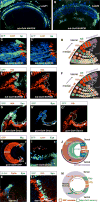
Figure 2. Lawf Neurons Are Generated in the Lamina Side of the OPC Neuroepithelium in Dpp Regions
(A and B) Randomly induced MARCM clones marked by tub-Gal4 label homogeneous groups of Lawf1 (A) or Lawf2 (B) neurons in adults. (C) Apterous (Ap, blue), expressed in half of medulla neurons (Li et al., 2013), is not expressed in Lawf2 Lim1+ neurons (Lim1, red) in a medulla NB clone. (D) Schematic representation of medulla and Lawf neurogenesis. Medulla NBs are generated from the medulla side of the neuroepithelium and progress through a tTF series beginning with Hth. Hth is also expressed in neuroepithelium cells before they become NBs. Lawf neurons are not generated by medulla NBs (dashed line outlines a medulla NB clone). (E) Clone from the lamina side of the OPC neuroepithelium generates progeny into the medulla neuron region. Progeny maintain Hth and Eya expression, confirming their Lawf identity, and migrate into the medulla neuropil. (F) Schematic representation as in (D) showing a neuroepithelial clone generating Lawf progeny (dashed line). (G) gcm-Gal4 G-trace where GFP marks Gal4 history. Lawf neurons (Eya+ and Hth+) originate from precursor cells in the lamina side of the neuroepithelium and migrate to the medulla (arrow shows direction of migration). (H) gcm-Gal4 drives expression on the lamina side of the OPC neuroepithelium of the main lamina region and dpp regions in the dorsal and ventral tips. The expression of gcm-Gal4 overlaps slightly with Wg regions. (I) Lawf1 cells are marked by the ventral-specific hh-Gal4 driving G-trace. Lawf2 cells (magenta) from the dorsal OPC are not marked by hh-Gal4 > G-trace. (J) Schematic representation of (H) and (I). (K) Eya+ Lawf precursor cells delaminate from the neuroepithelium region marked by dpp-lacZ (arrow shows the direction of delamination). (L) Lawf neurons in medulla marked by dpp-Gal4 driven G-trace. (M) Schematic representation of Lawf1 origin from the ventral Dpp (Gcm) region and Lawf2 origin from the dorsal Dpp (Gcm) region. See also Figure S2 and Movie S1.
Figure 3
Figure 3. Lawf Neurons, Lamina Epithelial, and Marginal Glia Share Common Progenitor Cells
(A) Schematic showing delaminating neuroepithelial cells in the GPC region giving rise to common progenitors (gray dashed line) that generate Lawf neurons and epithelial (eg) and marginal glia (mg; green dashed line) (B and C) Lawf neurons (Eya+, Elav+) and lamina eg and mg cells (Repo+) are labeled by 10C12-Gal4 driving UAS-GFP. (D) Progenitors marked by 10C12-Gal4-driven GFP generate both Lawfs (blue arrow) and Lamina glia (eg/mg; red arrow). Arrows indicate migratory routes. In the glia sub-lineage, Optix expression is turned on (D′), while Eya expression is reduced (D″). Arrowheads mark cells that coexpress Eya and Optix. (E) 10C12-Gal4 drives GFP in Eya+ progenitor cells delaminating from the neuroepithelium stained with anti-DE-cadherin (DECAD). Gray dashed line shows the location of the common progenitor cells. (F) Staining of the NB marker Dpn and neuropil marker DE-cadherin (DECAD) in the progenitor delamination region of neuroepithelium. Eya marks delaminating common progenitor cells. Gray dashed line shows the location of the common progenitor cells. (G) Ase and Pros staining in common progenitor cells (10C12-Gal4) indicate resemblance with GMCs. Pros is expressed when cells are still in the neuroepithelium, whereas Ase is expressed in common precursor cells after delamination. (H–J) Mitotic activity (PH3) in Lawf and glia sub-lineages. (H) Proliferating cells marked by PH3 staining in the neuroepithelium (arrowheads) and common progenitor cells (arrow). (I and I′) A number of Repo+ glial cells still maintain mitotic activity during migration. (J) Rare mitotic events are detected in the Lawf sub-lineage. (K) Model of Lawf and lamina glia (eg/mg) origin from common progenitor cells with their mitotic potential indicated. See also Figure S3 and Movies S1 and S2.
Figure 4
Figure 4. Live Imaging of 10C12>CD8GFP Reveals the Origin and Mitotic Potential of Lawf Neurons
(A) Diagram of L3 brains showing common progenitor cells (on the dorsal side), Lawf progenitors, and committed Lawfs and lamina glial cells along with their mitotic potential. Lawf and glial lineage migratory routes are indicated by arrows. (B–D) Still images captured from time-lapse two-photon imaging of the same late L3 10C12>CD8GFP eye-brain complex explant. 10C12-Gal4 drives GFP expression in all epithelial and marginal glia and in all Lawfs. The complete dorsal side of the lamina glial layers and Lawf cells are visible along with a small portion of the ventral region. The orientation of the optic lobe is labeled in (B.i) and matches (A). Lawfs and their precursors are marked by green dots and glial and their precursors are marked by red dots. Time is displayed in hours: minutes: seconds: milliseconds. Scale bar, 40 μm. (B.i and B.ii) A presumptive precursor in the most posterior dorsal region undergoes mitosis (arrowhead). This region is defined as the common progenitor region (dashed circle) and is populated by mitotically active cells (refer to Movie S2). (B.iii and B.iv) A restricted neuronal precursor destined to take on Lawf fate (green dot) migrates proximally from the common precursor region and (B.v) undergoes mitosis, (B.vi–B.viii) resulting in two Lawf cells, which begin to migrate tangentially. (C) A population of committed Lawfs (green dots) migrate tangentially over substantial distances from a starting point proximal to the common precursor pool. These cells appear to grow their axons during the course of migration but more prominently once they reach more anterior positions. (D.i–D.iv) A cell (red dot) migrates to join the lamina glial layers from the precursor region (dashed circle) along a route distinct from the Lawfs to enter the epithelial glial layer from the distal side. (D.v–D.xii) Two additional glia/glial precursors (D.v and D.viii) migrate shorter distances to join the presumptive marginal glial layer from the proximal side. Note that cells that are lost during the tracking process are changed to a lighter color and kept stationary. See also Movie S2.
Figure 5
Figure 5. Notch-Dependent Binary Fate Choice between Lawf and Lamina Glial Precursors
(A–C) 10C12-Gal4 marked MARCM clones (control and mutant) in the common progenitor cells close to the neuroepithelium, and in committed Lawf and glial lineages (D–F). (A) A control 10C12 Gal4 clone contains both Lawf (Eya+, blue) and Glia (Optix+, red) sub-lineages. (B) In a Su(H) mutant clone, which contains Lawfs (Eya+, blue), glial cells (Optix+, red) are not recovered. (C) In a numb mutant clone, glial cells (Optix+, red), but not Lawfs (Eya+), are recovered. (D) Control clone in a more anterior region of the optic lobe contains both Lawf and lamina eg/mg cells. (E) Su(H) mutant cells develop into Lawf neurons as they mature but never include glia. (F) Mature numb mutant cells develop into lamina glial cells. (G–K) Brains of Lobesi mutants (lateral view). Ventral photoreceptors do not develop in Lobesi. (G) Ventral lamina precursor cells (region between the dashed lines) fail to proliferate and generate lamina. (H) Maximum projection. In Lobesi mutants, the ventral lamina is absent and shows impaired ventral gliogenesis. Photoreceptor axons (Chaoptin, blue) and lamina glia (Optix, red) are only in the central and dorsal parts of the lamina (above the dashed line). (I) Ventral glia precursor cells form a small cluster of Optix+ cells squeezed into the lobula plug region (dashed circle). (J) These clustered glial precursor cells are also Eya+. (K) Lawf-specific markers are unaffected in Lobesi mutant. (L) Model for Notch dependent fate decision between Lawfs neurons and lamina glia (eg and mg).

References

    1. Apitz H, Salecker I. A region-specific neurogenesis mode requires migratory progenitors in the Drosophila visual system. Nat Neurosci. 2015;18:46–55. - PMC - PubMed
    1. Bayraktar OA, Doe CQ. Combinatorial temporal patterning in progenitors expands neural diversity. Nature. 2013;498:449–455. - PMC - PubMed
    1. Bayraktar OA, Boone JQ, Drummond ML, Doe CQ. Drosophila type II neuroblast lineages keep Prospero levels low to generate large clones that contribute to the adult brain central complex. Neural Dev. 2010;5:26. - PMC - PubMed
    1. Behnia R, Clark DA, Carter AG, Clandinin TR, Desplan C. Processing properties of ON and OFF pathways for Drosophila motion detection. Nature. 2014;512:427–430. - PMC - PubMed
    1. Bello BC, Izergina N, Caussinus E, Reichert H. Amplification of neural stem cell proliferation by intermediate progenitor cells in Drosophila brain development. Neural Dev. 2008;3:5. - PMC - PubMed

LinkOut - more resources