. 2016 Apr 26;15(4):692-699.
doi: 10.1016/j.celrep.2016.03.067. Epub 2016 Apr 14.

Improved Monosynaptic Neural Circuit Tracing Using Engineered Rabies Virus Glycoproteins

Affiliations

Improved Monosynaptic Neural Circuit Tracing Using Engineered Rabies Virus Glycoproteins

Euiseok J Kim et al. Cell Rep. .

Abstract

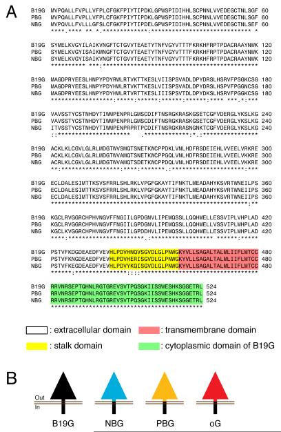
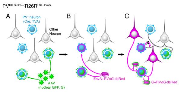
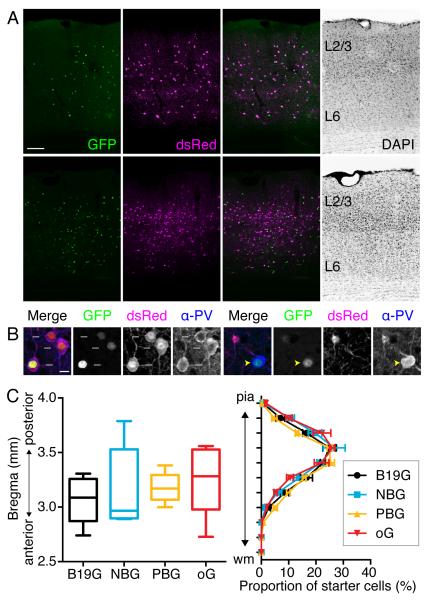
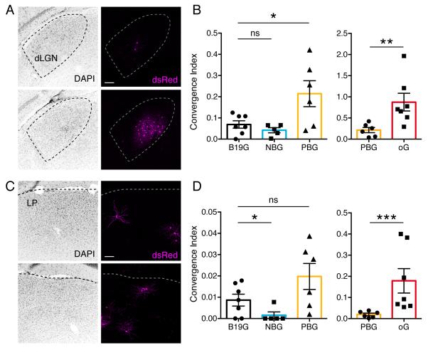
Monosynaptic rabies virus tracing is a unique and powerful tool used to identify neurons making direct presynaptic connections onto neurons of interest across the entire nervous system. Current methods utilize complementation of glycoprotein gene-deleted rabies of the SAD B19 strain with its glycoprotein, B19G, to mediate retrograde transsynaptic spread across a single synaptic step. In most conditions, this method labels only a fraction of input neurons and would thus benefit from improved efficiency of transsynaptic spread. Here, we report newly engineered glycoprotein variants to improve transsynaptic efficiency. Among them, oG (optimized glycoprotein) is a codon-optimized version of a chimeric glycoprotein consisting of the transmembrane/cytoplasmic domain of B19G and the extracellular domain of rabies Pasteur virus strain glycoprotein. We demonstrate that oG increases the tracing efficiency for long-distance input neurons up to 20-fold compared to B19G. oG-mediated rabies tracing will therefore allow identification and study of more complete monosynaptic input neural networks.


Figures

Figure 1
Figure 1. Rabies virus glycoprotein from B19 strain and its three engineered variants
(A) Multiple protein sequence alignment for B19G, PBG, and NBG. ’*’ indicates a single fully conserved amino acid residue. ’:’ indicates conservation between amino acid groups of strongly similar properties, whereas ’.’ indicates conservation between amino acid groups of weakly similar properties (the Gonnet PAM 250 matrix score >0.5 and =<0.5, respectively; (Sievers et al., 2011)). (B) Schematic representation illustrating glycoprotein from B19 strain and three other chimeric variants: NBG, PBG and oG. Yellow highlighted sequences indicate stalk domain, red indicate transmembrane domain, and green indicate cytoplasmic domain of glycoproteins.
Figure 2
Figure 2. Monosynaptic rabies virus tracing of inputs to PV interneurons
Schematic illustration of the scheme for virus infection and spread of rabies virus from PV interneurons in primary visual cortex (V1) of PVIRES-Cre/+;R26LSL-TVA/+ mouse. (A) PVIRES-Cre/+;R26LSL-TVA/+ mice were injected in V1 with AAV-FLEX-H2B-GFP-2A-GWPRE. All PV interneurons express TVA (blue) and those that are starter cells also express nuclear GFP (H2B-GFP) and rabies glycoprotein (G). (B) After two or three weeks, EnvA+RVdG-dsRed was injected into the same site in V1. EnvA+RVdG-dsRed infects TVA-expressing PV interneurons including starter cells. (C) After one week, monosynaptic input neurons to PV starter neurons were labeled with dsRed+ due to retrograde trans-synaptic spread of rabies virus.
Figure 3
Figure 3. PV starter neuron distributions in mouse V1
(A) Coronal sections of mouse V1 show starter neurons expressing both GFP from AAV-FLEX-H2B-GFP-2A-G and dsRed from EnvA+RVdG-dsRed. Top panels illustrate a mouse V1 section when B19G was used, whereas bottom panels illustrate use of oG. (B) Confocal images showing PV starter neurons (left panels, white arrows indicate GFP+ dsRed+ α-PV+ neurons) and PV+ non-starter neurons (right panels, yellow arrowheads indicate GFP+ α-PV+, but dsRed neurons) (C) Starter cell distributions. (left panel) the median position of starter neuron populations expressed as center of gravity along the anterior-posterior axis for each of the four experimental groups labeled according the glycoprotein used for transcomplementation. Values (mm) indicate distances posterior to bregma. Boxes extend from the 25th to 75th percentile. Whiskers indicate ranges from smallest to largest values. There are no statistical differences between groups (Kruskal-Wallis test with Dunn’s multiple comparisons test, p=0.5288). (right panel) Distributions of starter neuron proportions plotted according to distance from pia to white matter (wm) (Kruskal-Wallis test with Dunn’s multiple comparisons test, p=0.9992). The values are reported as mean ± SEM. Scale bars = 100 μm (A), 10 μm (B).
Figure 4
Figure 4. Efficiency of trans-synaptic spread following trans-complementation with different glycoproteins
Coronal sections showing dsRed+ monosynaptic input neurons in dLGN (A) and LP (C) to PV interneurons in V1 when using B19G (top panels) and oG (bottom panels). (B and D) Convergence indices for long-distance presynaptic inputs in dLGN (B) and LP (D) when using B19G, NBG, PBG, or oG rabies glycoproteins. Values are reported as mean ± SEM. Statistics were calculated from Mann-Whitney test for non-parametric comparisons. Significant differences between pairs are indicated by the p value. *p < 0.05, **p < 0.01, ***p < 0.001. Abbreviations are: dLGN, dorsal lateral geniculate nucleus; LP, lateral posterior thalamic nucleus. Scale bars = 100 μm (A, C).

References

    1. Callaway EM. Transneuronal circuit tracing with neurotropic viruses. Current Opinion in Neurobiology. 2008;18:617–623. - PMC - PubMed
    1. Callaway EM, Luo L. Monosynaptic Circuit Tracing with Glycoprotein-Deleted Rabies Viruses. J Neurosci. 2015;35:8979–8985. - PMC - PubMed
    1. Gerfen CR, Paletzki R, Heintz N. GENSAT BAC Cre-Recombinase Driver Lines to Study the Functional Organization of Cerebral Cortical and Basal Ganglia Circuits. Neuron. 2013;80:1368–1383. - PMC - PubMed
    1. Hippenmeyer S, Vrieseling E, Sigrist M, Portmann T, Laengle C, Ladle DR, Arber S. A developmental switch in the response of DRG neurons to ETS transcription factor signaling. PLoS Biol. 2005;3:e159. - PMC - PubMed
    1. Hirano M, Kato S, Kobayashi K, Okada T, Yaginuma H, Kobayashi K. Highly efficient retrograde gene transfer into motor neurons by a lentiviral vector pseudotyped with fusion glycoprotein. PLoS One. 2013;8:e75896. - PMC - PubMed