

Reproducibility of image-based computational models of intracranial aneurysm: a comparison between 3D rotational angiography, CT angiography and MR angiography
- PMID: 27150439
- PMCID: PMC4858827
- DOI: 10.1186/s12938-016-0163-4
Reproducibility of image-based computational models of intracranial aneurysm: a comparison between 3D rotational angiography, CT angiography and MR angiography
Abstract
Background: Reconstruction of patient-specific biomechanical model of intracranial aneurysm has been based on different imaging modalities. However, different imaging techniques may influence the model geometry and the computational fluid dynamics (CFD) simulation. The aim of this study is to evaluate the differences of the morphological and hemodynamic parameters in the computational models reconstructed from computed tomography angiography (CTA), magnetic resonance angiography (MRA) and 3D rotational angiography (3DRA).
Methods: Ten patients with cerebral aneurysms were enrolled in the study. MRA, CTA and 3DRA were performed on all patients. For each patient, three patient-specific models were reconstructed respectively based on the three sets of imaging data of the patient. CFD simulations were performed on each model. Model geometry and hemodynamic parameters were compared between the three models.
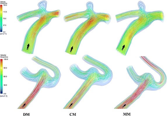
Results: In terms of morphological parameters, by comparing CTA based models (CM) and 3DRA based models (DM) which were treated as the "standard models", the aspect ratio had the minimum difference (Δ = 8.3 ± 1.72 %, P = 0.953) and the surface distance was 0.25 ± 0.07 mm. Meanwhile, by comparing MRA based models (MM) and DM, the size had the minimum difference (Δ = 6.6 ± 1.85 %, P = 0.683) and the surface distance was 0.36 ± 0.1 mm. In respect of hemodynamic parameters, all three models showed a similar distribution: low average WSS at the sack, high OSI at the body and high average WSSG at the neck. However, there was a large variation in the average WSS (Δ = 34 ± 5.13 % for CM, Δ = 40.6 ± 9.21 % for MM).
Conclusion: CTA and MRA have no significant differences in reproducing intracranial aneurysm geometry. The CFD results suggests there might be some significant differences in hemodynamic parameters between the three imaging-based models and this needs to be considered when interpreting the CFD results of different imaging-based models. If we only need to study the main flow patterns, three types of image-based model might be all suitable for patient-specific computational modeling studies.
Keywords: Aneurysm; Angiography; CFD; CTA; DSA; MRA; Reproducibility.
Figures


Similar articles
-
Assessing the Risk of Intracranial Aneurysm Rupture Using Morphological and Hemodynamic Biomarkers Evaluated from Magnetic Resonance Fluid Dynamics and Computational Fluid Dynamics.Magn Reson Med Sci. 2020 Dec 1;19(4):333-344. doi: 10.2463/mrms.mp.2019-0107. Epub 2020 Jan 17. Magn Reson Med Sci. 2020. PMID: 31956175 Free PMC article.
-
Reproducibility of image-based analysis of cerebral aneurysm geometry and hemodynamics: an in-vitro study of magnetic resonance imaging, computed tomography, and three-dimensional rotational angiography.J Neurol Surg A Cent Eur Neurosurg. 2013 Sep;74(5):294-302. doi: 10.1055/s-0033-1342937. Epub 2013 May 22. J Neurol Surg A Cent Eur Neurosurg. 2013. PMID: 23700168
-
Patient-specific computational hemodynamics of intracranial aneurysms from 3D rotational angiography and CT angiography: an in vivo reproducibility study.AJNR Am J Neuroradiol. 2011 Mar;32(3):581-6. doi: 10.3174/ajnr.A2306. Epub 2010 Dec 23. AJNR Am J Neuroradiol. 2011. PMID: 21183614 Free PMC article.
-
Computational Fluid Dynamics for Cerebral Aneurysms in Clinical Settings.Acta Neurochir Suppl. 2021;132:27-32. doi: 10.1007/978-3-030-63453-7_4. Acta Neurochir Suppl. 2021. PMID: 33973025 Review.
-
A review on the reliability of hemodynamic modeling in intracranial aneurysms: why computational fluid dynamics alone cannot solve the equation.Neurosurg Focus. 2019 Jul 1;47(1):E15. doi: 10.3171/2019.4.FOCUS19181. Neurosurg Focus. 2019. PMID: 31261119 Review.
Cited by
-
Volumetric surveillance of brain aneurysms: Pitfalls of MRA.Interv Neuroradiol. 2023 Oct;29(5):532-539. doi: 10.1177/15910199221100619. Epub 2022 May 12. Interv Neuroradiol. 2023. PMID: 35549745 Free PMC article.
-
Reproducibility of image-based computational models of intracranial aneurysm; methodological issue.Biomed Eng Online. 2016 Sep 16;15:109. doi: 10.1186/s12938-016-0223-9. Biomed Eng Online. 2016. PMID: 27637185 Free PMC article. No abstract available.
-
Quantitative and Qualitative Comparison of 4D-DSA with 3D-DSA Using Computational Fluid Dynamics Simulations in Cerebral Aneurysms.AJNR Am J Neuroradiol. 2019 Sep;40(9):1505-1510. doi: 10.3174/ajnr.A6172. AJNR Am J Neuroradiol. 2019. PMID: 31467234 Free PMC article.
-
Assessing the Risk of Intracranial Aneurysm Rupture Using Morphological and Hemodynamic Biomarkers Evaluated from Magnetic Resonance Fluid Dynamics and Computational Fluid Dynamics.Magn Reson Med Sci. 2020 Dec 1;19(4):333-344. doi: 10.2463/mrms.mp.2019-0107. Epub 2020 Jan 17. Magn Reson Med Sci. 2020. PMID: 31956175 Free PMC article.
-
Middle cerebral artery pressure laterality in patients with symptomatic ICA stenosis.PLoS One. 2021 Jan 8;16(1):e0245337. doi: 10.1371/journal.pone.0245337. eCollection 2021. PLoS One. 2021. PMID: 33417614 Free PMC article.
References
Publication types
MeSH terms
LinkOut - more resources
Full Text Sources
Other Literature Sources
Medical
Research Materials
Miscellaneous
