

Noncanonical connections between the subiculum and hippocampal CA1
- PMID: 27150503
- PMCID: PMC5050062
- DOI: 10.1002/cne.24024
Noncanonical connections between the subiculum and hippocampal CA1
Abstract
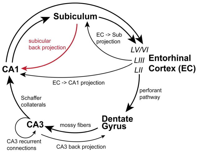
The hippocampal formation is traditionally viewed as having a feedforward, unidirectional circuit organization that promotes propagation of excitatory processes. While the substantial forward projection from hippocampal CA1 to the subiculum has been very well established, accumulating evidence supports the existence of a significant backprojection pathway comprised of both excitatory and inhibitory elements from the subiculum to CA1. Based on these recently updated anatomical connections, such a backprojection could serve to modulate information processing in hippocampal CA1. Here we review the published anatomical and physiological studies on the subiculum to CA1 backprojection, and present recent conclusive anatomical evidence for the presence of noncanonical subicular projections to CA1. New insights into this understudied pathway will improve our understanding of reciprocal CA1-subicular connections and guide future studies on how the subiculum interacts with CA1 to regulate hippocampal circuit activity and learning and memory behaviors. J. Comp. Neurol. 524:3666-3673, 2016. © 2016 The Authors The Journal of Comparative Neurology Published by Wiley Periodicals, Inc.
Keywords: back projection; genetic targeting; hippocampus; learning and memory; viral tracing.
© 2016 Wiley Periodicals, Inc.
Conflict of interest statement
Statement of conflict of interests All authors disclose no conflict of interests for this work.
Figures
Similar articles
-
Noncanonical projections to the hippocampal CA3 regulate spatial learning and memory by modulating the feedforward hippocampal trisynaptic pathway.PLoS Biol. 2021 Dec 20;19(12):e3001127. doi: 10.1371/journal.pbio.3001127. eCollection 2021 Dec. PLoS Biol. 2021. PMID: 34928938 Free PMC article.
-
Hippocampal CA3 inhibitory neurons receive extensive noncanonical synaptic inputs from CA1 and subicular complex.J Comp Neurol. 2023 Sep;531(13):1333-1347. doi: 10.1002/cne.25510. Epub 2023 Jun 13. J Comp Neurol. 2023. PMID: 37312626 Free PMC article.
-
Opposing and Complementary Topographic Connectivity Gradients Revealed by Quantitative Analysis of Canonical and Noncanonical Hippocampal CA1 Inputs.eNeuro. 2018 Jan 30;5(1):ENEURO.0322-17.2018. doi: 10.1523/ENEURO.0322-17.2018. eCollection 2018 Jan-Feb. eNeuro. 2018. PMID: 29387780 Free PMC article.
-
Constraints on hippocampal processing imposed by the connectivity between CA1, subiculum and subicular targets.Behav Brain Res. 2006 Nov 11;174(2):265-71. doi: 10.1016/j.bbr.2006.06.014. Epub 2006 Jul 21. Behav Brain Res. 2006. PMID: 16859763 Review.
-
The subiculum: the heart of the extended hippocampal system.Prog Brain Res. 2015;219:65-82. doi: 10.1016/bs.pbr.2015.03.003. Epub 2015 May 14. Prog Brain Res. 2015. PMID: 26072234 Review.
Cited by
-
Alteration of Neural Pathways and Its Implications in Alzheimer's Disease.Biomedicines. 2022 Apr 4;10(4):845. doi: 10.3390/biomedicines10040845. Biomedicines. 2022. PMID: 35453595 Free PMC article. Review.
-
Subiculum as a generator of sharp wave-ripples in the rodent hippocampus.Cell Rep. 2021 Apr 20;35(3):109021. doi: 10.1016/j.celrep.2021.109021. Cell Rep. 2021. PMID: 33882307 Free PMC article.
-
Noncanonical projections to the hippocampal CA3 regulate spatial learning and memory by modulating the feedforward hippocampal trisynaptic pathway.PLoS Biol. 2021 Dec 20;19(12):e3001127. doi: 10.1371/journal.pbio.3001127. eCollection 2021 Dec. PLoS Biol. 2021. PMID: 34928938 Free PMC article.
-
Differential expression of microRNAs in the hippocampi of male and female rodents after chronic alcohol administration.Biol Sex Differ. 2020 Nov 23;11(1):65. doi: 10.1186/s13293-020-00342-3. Biol Sex Differ. 2020. PMID: 33228793 Free PMC article.
-
CA1-projecting subiculum neurons facilitate object-place learning.Nat Neurosci. 2019 Nov;22(11):1857-1870. doi: 10.1038/s41593-019-0496-y. Epub 2019 Sep 23. Nat Neurosci. 2019. PMID: 31548723 Free PMC article.
References
-
- Amaral DG. Emerging principles of intrinsic hippocampal organization. Current opinion in neurobiology. 1993;3:225–229. - PubMed
-
- Amaral DG, Dolorfo C, Alvarez-Royo P. Organization of CA1 projections to the subiculum: a PHA-L analysis in the rat. Hippocampus. 1991;1:415–435. - PubMed
-
- Amaral DG, Witter MP. The three-dimensional organization of the hippocampal formation: a review of anatomical data. Neuroscience. 1989;31:571–591. - PubMed
-
- Berger TW, Swanson GW, Milner TA, Lynch GS, Thompson RF. Reciprocal anatomical connections between hippocampus and subiculum in the rabbit evidence for subicular innervation of regio superior. Brain Res. 1980;183:265–276. - PubMed
Publication types
MeSH terms
Grants and funding
LinkOut - more resources
Full Text Sources
Other Literature Sources
Miscellaneous
