

Perfusion-related stimuli for compensatory lung growth following pneumonectomy
- PMID: 27150830
- PMCID: PMC4967253
- DOI: 10.1152/japplphysiol.00297.2016
Perfusion-related stimuli for compensatory lung growth following pneumonectomy
Abstract
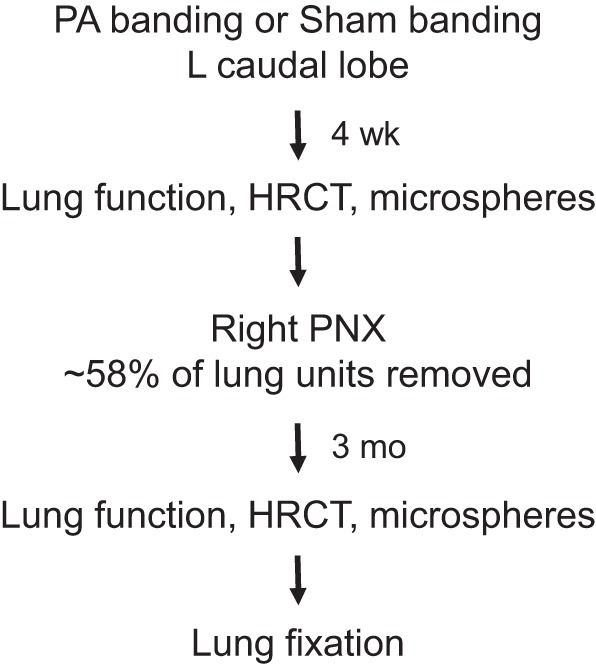
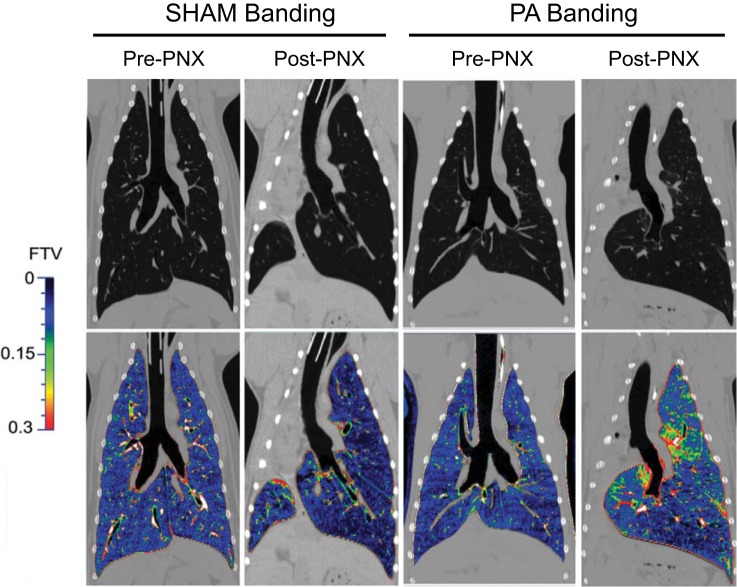
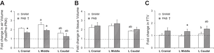
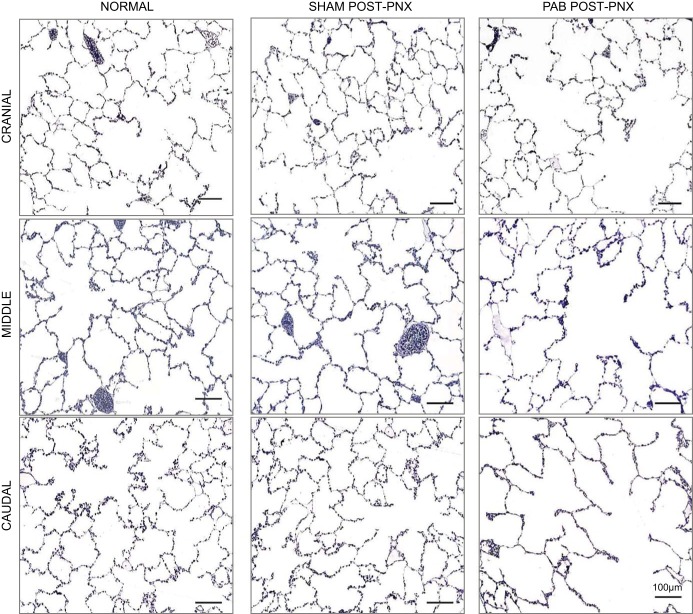
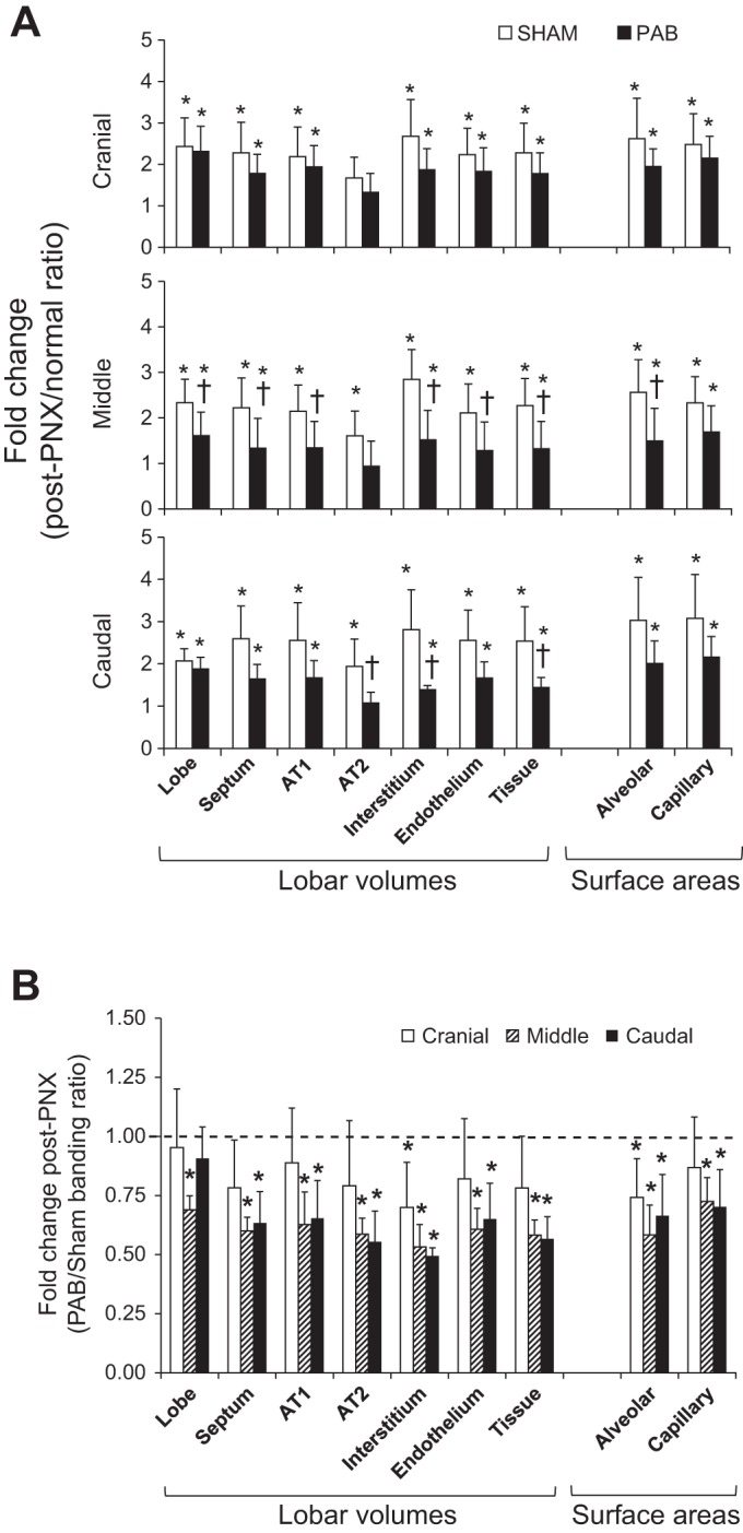
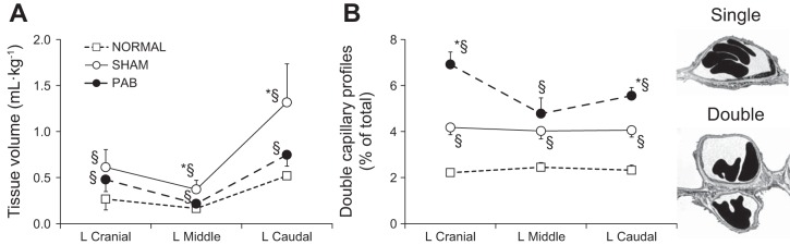
Following pneumonectomy (PNX), two separate mechanical forces act on the remaining lung: parenchymal stress caused by lung expansion, and microvascular distension and shear caused by increased perfusion. We previously showed that parenchymal stress and strain explain approximately one-half of overall compensation; the remainder was presumptively attributed to perfusion-related factors. In this study, we directly tested the hypothesis that perturbation of regional pulmonary perfusion modulates post-PNX lung growth. Adult canines underwent banding of the pulmonary artery (PAB) to the left caudal (LCa) lobe, which caused a reduction in basal perfusion to LCa lobe without preventing the subsequent increase in its perfusion following right PNX while simultaneously exaggerating the post-PNX increase in perfusion to the unbanded lobes, thereby creating differential perfusion changes between banded and unbanded lobes. Control animals underwent sham pulmonary artery banding followed by right PNX. Pulmonary function, regional pulmonary perfusion, and high-resolution computed tomography of the chest were analyzed pre-PNX and 3-mo post-PNX. Terminally, the remaining lobes were fixed for detailed morphometric analysis. Results were compared with corresponding lobes in two control (Sham banding and normal unoperated) groups. PAB impaired the indices of post-PNX extravascular alveolar tissue growth by up to 50% in all remaining lobes. PAB enhanced the expected post-PNX increase in alveolar capillary formation, measured by the prevalence of double-capillary profiles, in both unbanded and banded lobes. We conclude that perfusion distribution provides major stimuli for post-PNX compensatory lung growth independent of the stimuli provided by lung expansion and parenchymal stress and strain.
Keywords: alveolar angiogenesis; lung resection; lung structure and function; pulmonary artery banding; pulmonary blood flow.
Copyright © 2016 the American Physiological Society.
Figures
Similar articles
-
Separating in vivo mechanical stimuli for postpneumonectomy compensation: physiological assessment.J Appl Physiol (1985). 2013 Jan 1;114(1):99-106. doi: 10.1152/japplphysiol.01213.2012. Epub 2012 Oct 25. J Appl Physiol (1985). 2013. PMID: 23104695 Free PMC article.
-
Regional lung growth following pneumonectomy assessed by computed tomography.J Appl Physiol (1985). 2004 Oct;97(4):1567-74; discussion 1549. doi: 10.1152/japplphysiol.00396.2004. Epub 2004 Jun 18. J Appl Physiol (1985). 2004. PMID: 15208290
-
Separating in vivo mechanical stimuli for postpneumonectomy compensation: imaging and ultrastructural assessment.J Appl Physiol (1985). 2013 Apr;114(8):961-70. doi: 10.1152/japplphysiol.01394.2012. Epub 2013 Jan 17. J Appl Physiol (1985). 2013. PMID: 23329819 Free PMC article.
-
Further examination of alveolar septal adaptation to left pneumonectomy in the adult lung.Respir Physiol Neurobiol. 2006 Apr 28;151(2-3):167-77. doi: 10.1016/j.resp.2006.01.013. Epub 2006 Mar 24. Respir Physiol Neurobiol. 2006. PMID: 16563882 Review.
-
The pneumonectomy model of compensatory lung growth: insights into lung regeneration.Pharmacol Ther. 2014 May;142(2):196-205. doi: 10.1016/j.pharmthera.2013.12.006. Epub 2013 Dec 12. Pharmacol Ther. 2014. PMID: 24333263 Review.
Cited by
-
Inhalational delivery of induced pluripotent stem cell secretome improves postpneumonectomy lung structure and function.J Appl Physiol (1985). 2020 Nov 1;129(5):1051-1061. doi: 10.1152/japplphysiol.00205.2020. Epub 2020 Sep 10. J Appl Physiol (1985). 2020. PMID: 32909918 Free PMC article.
-
In vivo imaging of canine lung deformation: effects of posture, pneumonectomy, and inhaled erythropoietin.J Appl Physiol (1985). 2020 May 1;128(5):1093-1105. doi: 10.1152/japplphysiol.00647.2019. Epub 2020 Jan 16. J Appl Physiol (1985). 2020. PMID: 31944885 Free PMC article.
-
No compensatory lung growth after resection in a one-year follow-up cohort of patients with lung cancer.J Thorac Dis. 2017 Oct;9(10):3938-3945. doi: 10.21037/jtd.2017.08.135. J Thorac Dis. 2017. PMID: 29268404 Free PMC article.
-
Blood flow-induced angiocrine signals promote organ growth and regeneration.Bioessays. 2025 Feb;47(2):e2400207. doi: 10.1002/bies.202400207. Epub 2024 Nov 11. Bioessays. 2025. PMID: 39529434 Free PMC article. Review.
-
Erythropoietin inhalation enhances adult canine alveolar-capillary formation following pneumonectomy.Am J Physiol Lung Cell Mol Physiol. 2019 May 1;316(5):L936-L945. doi: 10.1152/ajplung.00504.2018. Epub 2019 Feb 20. Am J Physiol Lung Cell Mol Physiol. 2019. PMID: 30785346 Free PMC article.
References
-
- Abdulnour RE, Peng X, Finigan JH, Han EJ, Hasan EJ, Birukov KG, Reddy SP, Watkins JE III, Kayyali US, Garcia JG, Tuder RM, Hassoun PM. Mechanical stress activates xanthine oxidoreductase through MAP kinase-dependent pathways. Am J Physiol Lung Cell Mol Physiol 291: L345–L353, 2006. - PubMed
-
- Berger LC, Burri PH. Timing of the quantitative recovery in the regenerating rat lung. Am Rev Respir Dis 132: 777–783, 1985. - PubMed
-
- Burri PH. Structural aspects of postnatal lung development: alveolar formation and growth. Biol Neonate 89: 313–322, 2006. - PubMed
-
- Burri PH, Pfrunder HB, Berger LC. Reactive changes in pulmonary parenchyma after bilobectomy: a scanning electron microscopic investigation. Exp Lung Res 4: 11–28, 1982. - PubMed
-
- Carlin JI, Hsia CC, Cassidy SS, Ramanathan M, Clifford PS, Johnson RL Jr. Recruitment of lung diffusing capacity with exercise before and after pneumonectomy in dogs. J Appl Physiol 70: 135–142, 1991. - PubMed
Publication types
MeSH terms
Grants and funding
LinkOut - more resources
Full Text Sources
Other Literature Sources
Research Materials