Review
. 2016 Sep;65(3):608-17.
doi: 10.1016/j.jhep.2016.04.018. Epub 2016 May 2.

Sinusoidal communication in liver fibrosis and regeneration

Affiliations
Review

Sinusoidal communication in liver fibrosis and regeneration

Giusi Marrone et al. J Hepatol. 2016 Sep.

Abstract

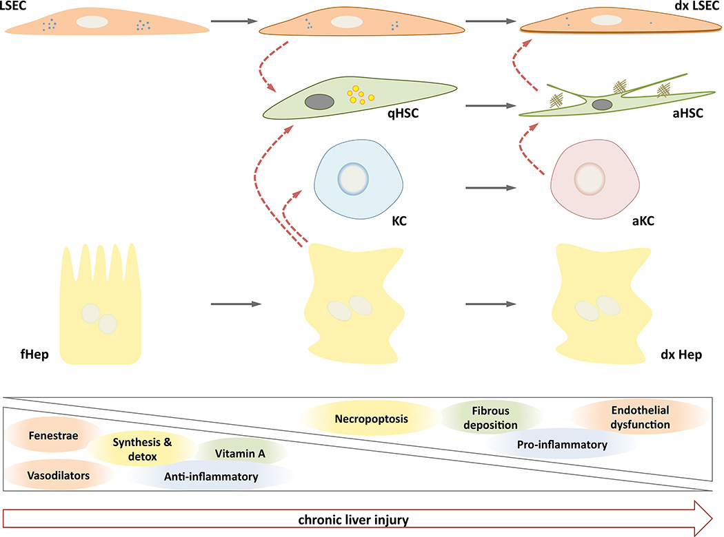
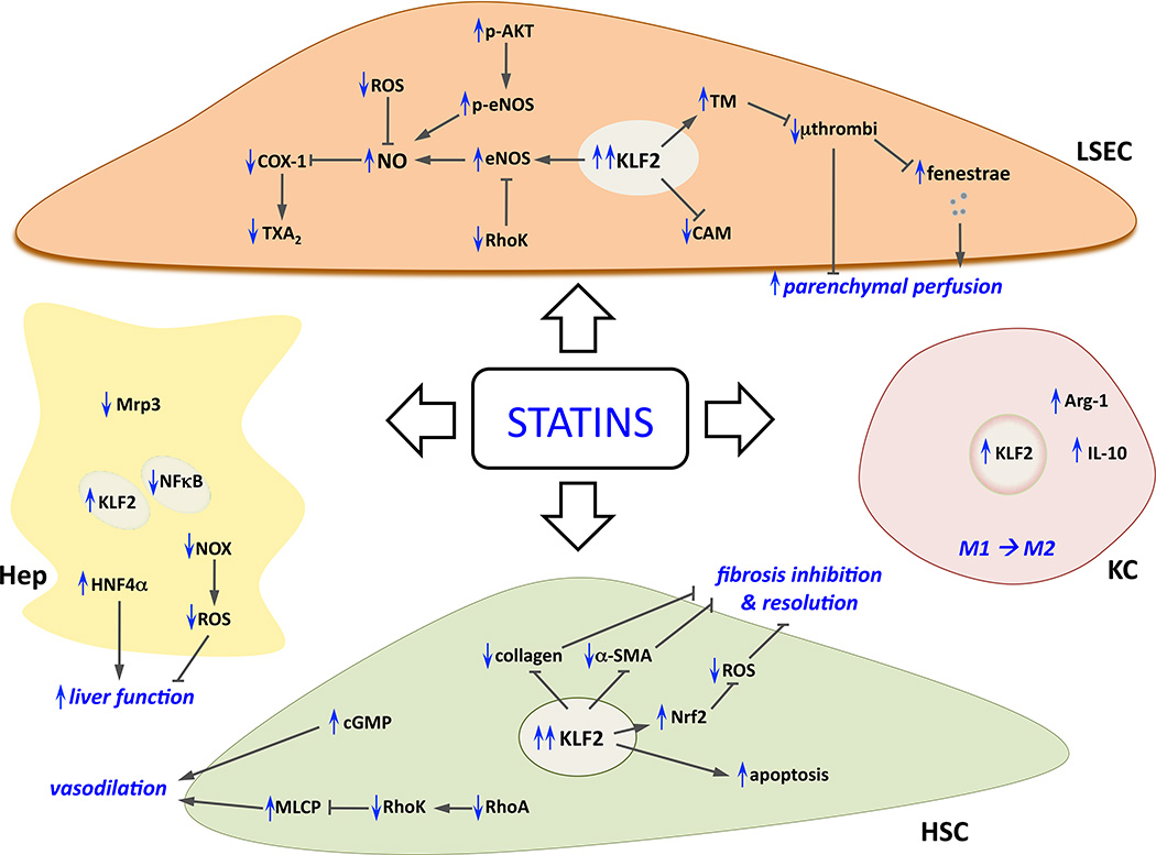
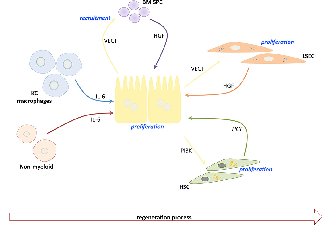
Cellular crosstalk is a process through which a message is transmitted within an individual cell (intracellular crosstalk) or between different cells (intercellular crosstalk). Intercellular crosstalk within the liver microenvironment is critical for the maintenance of normal hepatic functions and for cells survival. Hepatic cells are closely connected to each other, work in synergy, and produce molecules that modulate their differentiation and activity. This review summarises the current knowledge regarding paracrine communication networks in parenchymal and non-parenchymal cells in liver fibrosis due to chronic injury, and regeneration after partial hepatectomy.

Keywords: Cirrhosis; HSC; Hepatic stellate cells; Hepatocytes; Ischemia/reperfusion; Kupffer cells; LSEC; Liver sinusoidal endothelial cells; Portal hypertension; Regeneration; Transplantation.


Conflict of interest statement

None to declare.

Figures

Figure 1
Figure 1. Sinusoidal crosstalk during chronic liver injury
Initial de-regulations in functional Hepatocytes (fHep) and Liver Sinusoidal Endothelial Cells (LSEC) due to liver damage lead to complex paracrine interactions (red dotted arrows) with quiescent Hepatic Stellate Cells (qHSC) and Kupffer Cells (KC), ultimately creating a dysfunctional sinusoidal microenvironment composed by dysfunctional LSEC (dxLSEC), activated HSC (aHSC), activated KC (aKC) and dysfunctional and necroptotic hepatocytes (dxHep). Phenotypic characteristics of each cell type are progressively lost during the progression of the liver disease, and new pathologic properties appear.
Figure 2
Figure 2. Effects of statins on sinusoidal cells
Summary of cellular and molecular mechanisms underlying the beneficial effects of statins in liver cells (in blue, modifications due to statins administration). Please note that to simplify the figure paracrine interactions between statin-improved cells are intentionally omitted, complete explanation can be found along the text. α-SMA; smooth muscle actin alpha; Arg-1, arginase-1; CAM, cell adhesion molecules; cGMP, cyclic guanosine monophosphate; COX-1, cyclooxygenase-1; eNOS, endothelial nitric oxide synthase; HNF4α, hepatocyte nuclear factor 4 alpha; KLF2, Kruppel-like factor 2; MLCP, myosin light chain phosphatase; Mrp3, ATP-binding cassette sub-family C member 3; NFkB, nuclear factor kappa-light-chain-enhancer of activated B cells; NO, nitric oxide; NOX, NADPH oxidase; Nrf2, nuclear factor (erythroid-derived 2)-like 2; ROS, radical oxygen species; TM, thrombomodulin; TXA2, thromboxane A2.
Figure 3
Figure 3. Sinusoidal crosstalk during liver regeneration
Upon hepatectomy, initial signals (mainly IL-6) from Kupffer cells (KC), macrophages and other non-myeloid cells lead to the first burst of hepatocytes proliferation, which in turn will produce vascular endothelial growth factor (VEGF) to recruit bone marrow-derived sinusoidal progenitor cells (BM SPC) as a key source of hepatocyte growth factor (HGF). Latterly, hepatic stellate cells (HSC) and liver sinusoidal endothelial cells (LSEC) contribute to the regeneration process.

References

    1. Braet F, Wisse E. Structural and functional aspects of liver sinusoidal endothelial cell fenestrae: a review. Comp Hepatol. 2002;1:1. - PMC - PubMed
    1. Cogger VC, Warren A, Fraser R, Ngu M, McLean AJ, le Couteur DG. Hepatic sinusoidal pseudocapillarization with aging in the non-human primate. Exp Gerontol. 2003;38:1101–1107. - PubMed
    1. McLean AJ, Cogger VC, Chong GC, Warren A, Markus AM, Dahlstrom JE, et al. Age-related pseudocapillarization of the human liver. J Pathol. 2003;200:112–117. - PubMed
    1. Schaffner F, Poper H. Capillarization of hepatic sinusoids in man. Gastroenterology. 1963;44:239–242. 239–242. - PubMed
    1. Urashima S, Tsutsumi m, Nakase K, Wang JS, Takada A. Studies on capillarization of the hepatic sinusoids in alcoholic liver disease. Alcohol Alcohol Suppl. 1993;1B:77–84. 77–84. - PubMed