Review
. 2016 Sep;73(18):3535-53.
doi: 10.1007/s00018-016-2260-8. Epub 2016 May 5.

Adherens Junction and E-Cadherin complex regulation by epithelial polarity

Affiliations
Review

Adherens Junction and E-Cadherin complex regulation by epithelial polarity

Peter Coopman et al. Cell Mol Life Sci. 2016 Sep.

Abstract

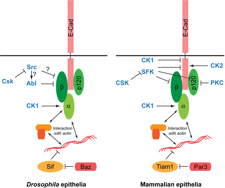
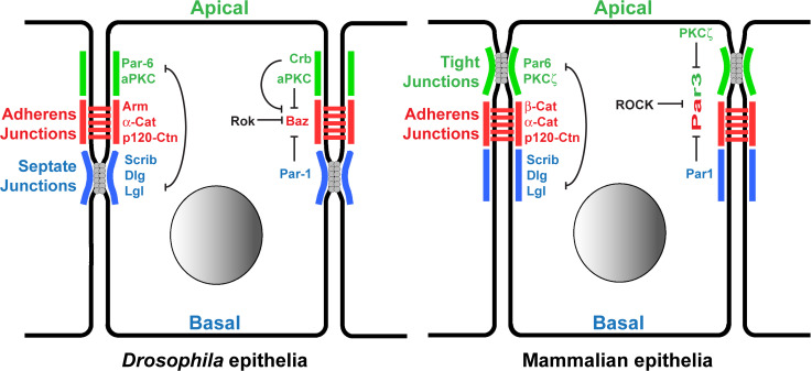
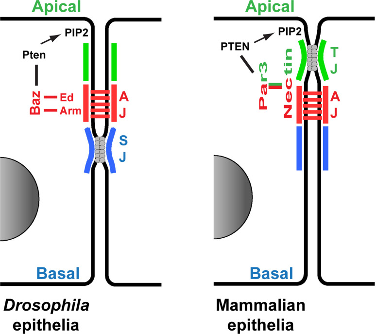
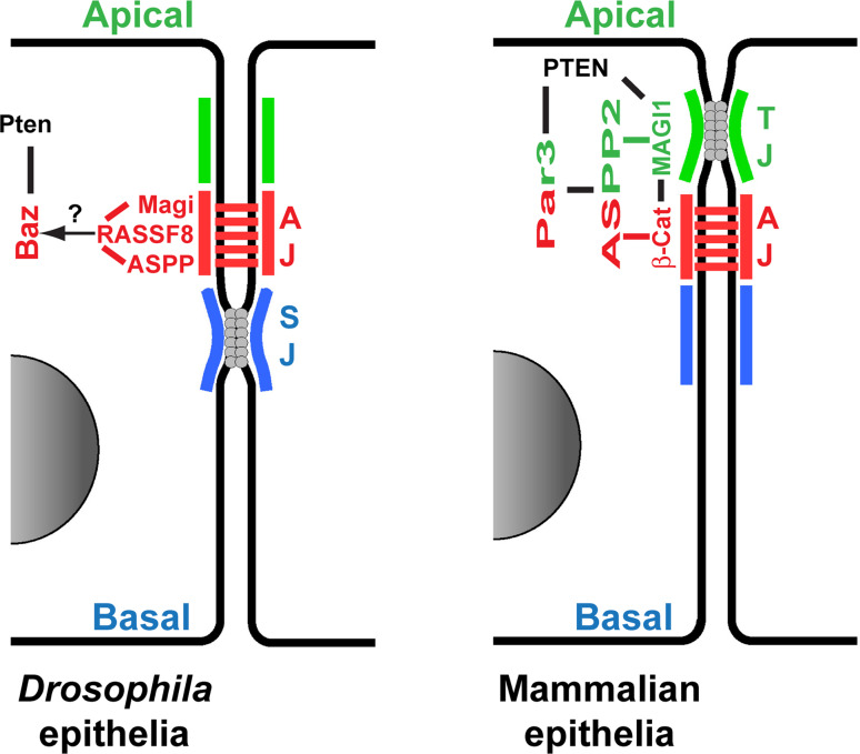
E-Cadherin-based Adherens Junctions (AJs) are a defining feature of all epithelial sheets. Through the homophilic association of E-Cadherin molecules expressed on neighboring cells, they ensure intercellular adhesion amongst epithelial cells, and regulate many key aspects of epithelial biology. While their adhesive role requires these structures to remain stable, AJs are also extremely plastic. This plasticity allows for the adaptation of the cell to its changing environment: changes in neighbors after cell division, cell death, or cell movement, and changes in cell shape during differentiation. In this review we focus on the recent advances highlighting the critical role of the apico-basal polarity machinery, and in particular of the Par3/Bazooka scaffold, in the regulation and remodeling of AJs. We propose that by regulating key phosphorylation events on the core E-Cadherin complex components, Par3 and epithelial polarity promote meta-stable protein complexes governing the correct formation, localization, and functioning of AJ.

Keywords: Adherens Junctions; E-Cadherin; Epithelial polarity; Magi scaffolds; Par3; Remodeling.


Figures

Fig. 1
Fig. 1
Core E-Cadherin/Catenin complexes at Adherens Junctions and their regulation by phosphorylation. Shown is a simplified comparison of the Drosophila (left) and mammalian (right) Adherens Junction (AJ) complexes and key kinases (in bold and in blue) regulating the association and stability between E-Cadherin and the different catenins. Arrows represent phosphorylations with an activating function (e.g., stabilization of complexes and AJs); bars represent inhibitory phosphorylations
Fig. 2
Fig. 2
Apical restriction of Par3/Baz localization by inhibitory kinases. Structures of epithelial cells in Drosophila (left) and in mammals (right), with the different protein orthologues implicated in Par3/Baz restriction, are presented. AJs, (shown in red) are localized apically at the interface between the apical and basal membranous domains characterized by antagonizing protein complexes: the apical Par6/PKCζ (green), and the baso-lateral Dlg/Lgl/Scrib/Par1 (blue). Par3 accumulates at the level of AJs due to a double restriction by the apical kinase PKCζ and by the basal kinase Par1. The domains of these two kinases stay segregated due to the mutual exclusion of the apical (green) and baso-lateral (blue) protein complex networks. Par3 levels are further modulated along the circumference of the AJ domain by the antagonizing Rho-Kinase (ROCK)
Fig. 3
Fig. 3
Adherens Junction retention of Par3/Baz. Comparison between the Drosophila (left) and mammalian (right) mechanisms to anchor Par3/Baz at AJs. Lines (black, red and green) indicate physical interaction. PTEN promotes the production of PIP2 phospholipids, a major component of the plasma membrane, anchoring Par3/Baz at the membrane
Fig. 4
Fig. 4
A MAGI-ASPP network to localize Par3/Baz at the Adherens Junction. Comparison between the Drosophila (left) and mammalian (right) emerging role of the MAGI-ASPP network in AJ regulation. Lines (black, red and green) indicate physical interaction. Further work on this network will allow to clarify the exact localizations and interactions between the different members of the complexes, and between the MAGI-ASPP network and the other factors regulating E-Cad complexes and Par3 localization presented in Figs. 1, 2, and 3

References

    1. Bryant DM, Mostov KE. From cells to organs: building polarized tissue. Nat Rev Mol Cell Biol. 2008;9:887–901. doi: 10.1038/nrm2523. - DOI - PMC - PubMed
    1. Goldstein B, Macara IG. The PAR proteins: fundamental players in animal cell polarization. Dev Cell. 2007;13:609–622. doi: 10.1016/j.devcel.2007.10.007. - DOI - PMC - PubMed
    1. St Johnston D, Ahringer J. Cell polarity in eggs and epithelia: parallels and diversity. Cell. 2010;141:757–774. doi: 10.1016/j.cell.2010.05.011. - DOI - PubMed
    1. Tepass U. The apical polarity protein network in Drosophila epithelial cells: regulation of polarity, junctions, morphogenesis, cell growth, and survival. Annu Rev Cell Dev Biol. 2012;28:655–685. doi: 10.1146/annurev-cellbio-092910-154033. - DOI - PubMed
    1. Nelson WJ, Dickinson DJ, Weis WI. Roles of cadherins and catenins in cell-cell adhesion and epithelial cell polarity. Prog Mol Biol Transl Sci. 2013;116:3–23. doi: 10.1016/B978-0-12-394311-8.00001-7. - DOI - PubMed

Publication types