. 2016 May 6;352(6286):725-9.
doi: 10.1126/science.aad5081. Epub 2016 May 5.

Femtosecond structural dynamics drives the trans/cis isomerization in photoactive yellow protein

Affiliations

Femtosecond structural dynamics drives the trans/cis isomerization in photoactive yellow protein

Kanupriya Pande et al. Science. .

Abstract

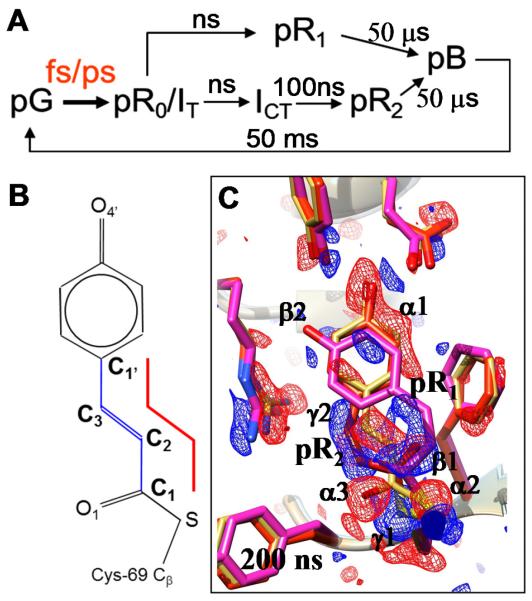
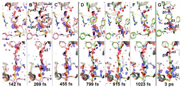
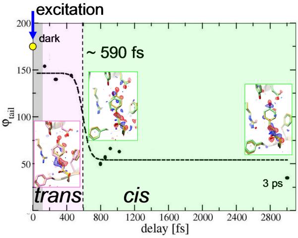
A variety of organisms have evolved mechanisms to detect and respond to light, in which the response is mediated by protein structural changes after photon absorption. The initial step is often the photoisomerization of a conjugated chromophore. Isomerization occurs on ultrafast time scales and is substantially influenced by the chromophore environment. Here we identify structural changes associated with the earliest steps in the trans-to-cis isomerization of the chromophore in photoactive yellow protein. Femtosecond hard x-ray pulses emitted by the Linac Coherent Light Source were used to conduct time-resolved serial femtosecond crystallography on photoactive yellow protein microcrystals over a time range from 100 femtoseconds to 3 picoseconds to determine the structural dynamics of the photoisomerization reaction.


Figures

Figure 1
Figure 1
A. The PYP photocycle from the perspective of a time-resolved crystallographer. Approximate time scales are given. The fs/ps time scale (in red) is structurally charted in this paper. B. The chemical structure of the pCA chromophore. The red line marks the four atoms that define the torsional angle ϕtail about the C2=C3 bond. C. Results of the positive control experiment at a 200 ns time delay. Reaction initiated by fs laser pulses. Negative (red) and positive (blue) DED features on the −3σ/3σ level. A mixture of the pR1 (magenta) and pR2 (red) structures is present. Main signature of pR1: features β1 and β2. Main signature for pR2: features γ1 and γ2. Structure of PYPref (dark) in
Figure 2
Figure 2
Trans to Cis isomerization in PYP. Weighted DED maps in red (−3 σ) and blue (3σ); front (upper) and side view (lower). Each map is prepared from about the same number of diffraction patterns, except the 3 ps map (see Tab. S1 B-C). The reference, dark structure is shown in yellow throughout; structures before the transition and still on the electronic excited state PES are shown in pink; structures after the transition and on the electronic ground state PES are shown in light green. Important negative difference density features are denoted α, positive features as β in panels B and G. Pronounced structural changes are marked by arrows. A-C: time-delays before the transition. A. Twisted trans at 142 fs, ϕtail 154°. B. Twisted trans at 269 fs, ϕtail 140° some important residues are marked; dotted lines in B: hydrogen bond of the ring hydroxyl to Glu46 and Tyr42. C. Twisted trans at 455 fs, ϕtail 144°; dotted line in C: direction of C2=C3 double bond. D-G: time delays and chromophore configuration after the transition. D. Early cis at 799 fs, ϕtail 50°. E. Early cis at 915 fs; dotted line in E: direction of C2=C3 double bond. F. Early cis at 1023 fs; for E and F ϕtail ~65°. G. 3 ps delay; dashed line: direction of C2=C3 double bond, feature β1; ϕtail is 35°.
Figure 3
Figure 3
Trans to cis isomerization in PYP. Pink: twisted trans on excited state PES; light green: cis on ground state PES. Torsional angle ϕtail (solid spheres) from structural refinement at various delays (see also Tab. S3). Gray region: not time-resolved. Dashed line: fit with eqn. S2, with a transition time of about 590 fs (see also Fig. S2). Inserts: structures of PYPfast (pink), PYPslow and PYP3ps (light green), and dark state structure PYPref in yellow. Difference electron density in red (−3σ) and blue (3σ).

References

    1. Wald G. Molecular basis of visual excitation. Science. 1968;162:230–239. - PubMed
    1. Mizutani Y, Kitagawa T. Direct observation of cooling of heme upon photodissociation of carbonmonoxy myoglobin. Science. 1997;278:443–446. - PubMed
    1. Nakamura R, Hamada N, Ichida H, Tokunaga F, Kanematsu Y. Coherent oscillations in ultrafast fluorescence of photoactive yellow protein. The Journal of chemical physics. 2007;127:215102. doi: 10.1063/1.2802297. published online EpubDec 7. - DOI - PubMed
    1. Champion PM. Chemistry. Following the flow of energy in biomolecules. Science. 2005;310:980–982. doi: 10.1126/science.1120280. published online EpubNov 11. - DOI - PMC - PubMed
    1. Lincoln CN, Fitzpatrick AE, van Thor JJ. Photoisomerisation quantum yield and non-linear cross-sections with femtosecond excitation of the photoactive yellow protein. Physical chemistry chemical physics : PCCP. 2012;14:15752–15764. doi: 10.1039/c2cp41718a. published online EpubDec 5. - DOI - PubMed

Publication types

Substances