Case Reports
. 2016 Aug;5(8):1098-105.
doi: 10.5966/sctm.2015-0358. Epub 2016 May 5.

Personalized Stem Cell Therapy to Correct Corneal Defects Due to a Unique Homozygous-Heterozygous Mosaicism of Ectrodactyly-Ectodermal Dysplasia-Clefting Syndrome

Affiliations
Case Reports

Personalized Stem Cell Therapy to Correct Corneal Defects Due to a Unique Homozygous-Heterozygous Mosaicism of Ectrodactyly-Ectodermal Dysplasia-Clefting Syndrome

Vanessa Barbaro et al. Stem Cells Transl Med. 2016 Aug.

Abstract

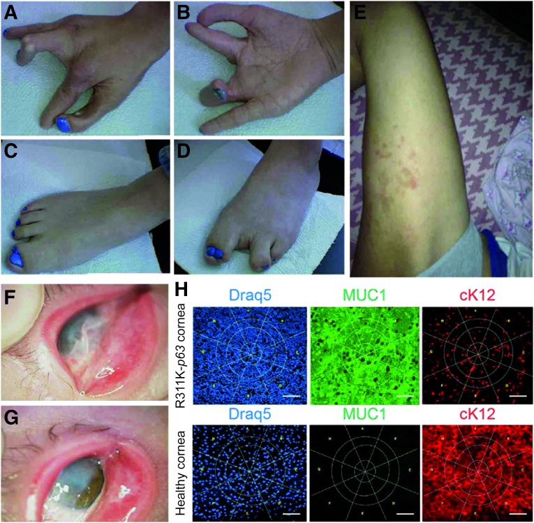
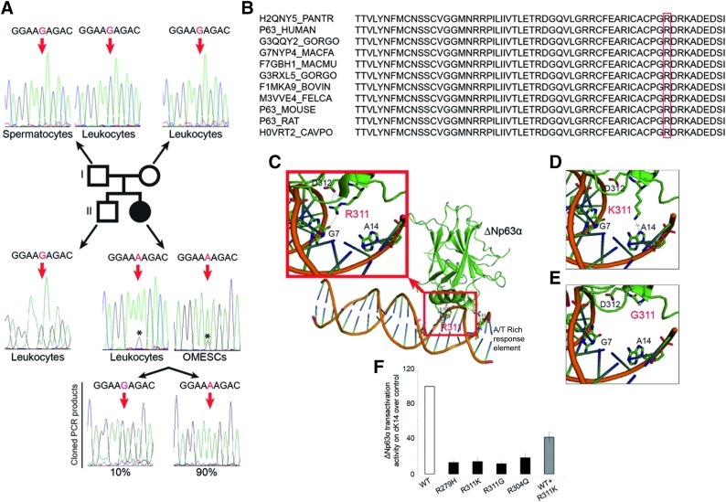
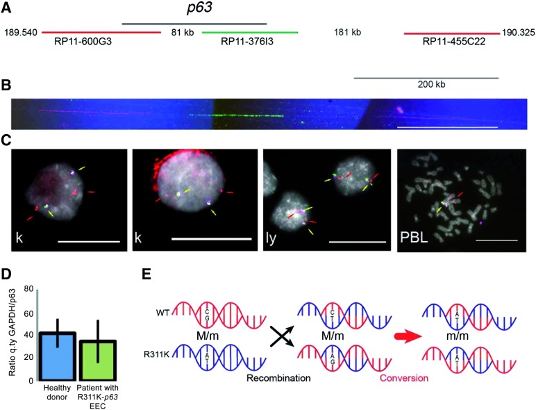
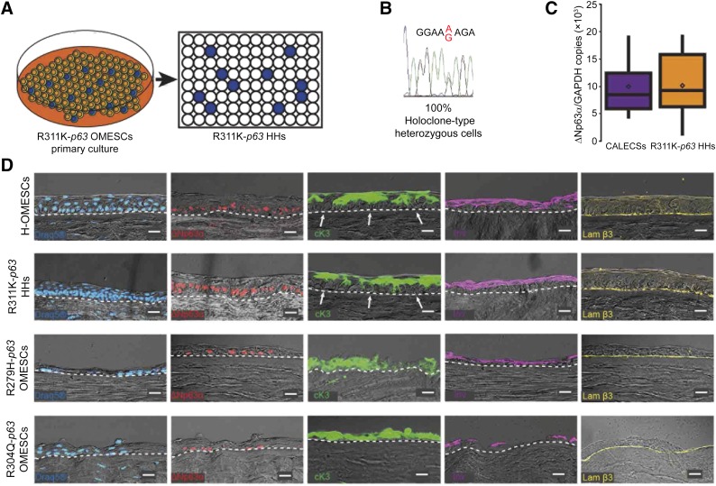
: Ectrodactyly-ectodermal dysplasia-clefting (EEC) syndrome is a rare autosomal dominant disease caused by mutations in the p63 gene. To date, approximately 40 different p63 mutations have been identified, all heterozygous. No definitive treatments are available to counteract and resolve the progressive corneal degeneration due to a premature aging of limbal epithelial stem cells. Here, we describe a unique case of a young female patient, aged 18 years, with EEC and corneal dysfunction, who was, surprisingly, homozygous for a novel and de novo R311K missense mutation in the p63 gene. A detailed analysis of the degree of somatic mosaicism in leukocytes from peripheral blood and oral mucosal epithelial stem cells (OMESCs) from biopsies of buccal mucosa showed that approximately 80% were homozygous mutant cells and 20% were heterozygous. Cytogenetic and molecular analyses excluded genomic alterations, thus suggesting a de novo mutation followed by an allelic gene conversion of the wild-type allele by de novo mutant allele as a possible mechanism to explain the homozygous condition. R311K-p63 OMESCs were expanded in vitro and heterozygous holoclones selected following clonal analysis. These R311K-p63 OMESCs were able to generate well-organized and stratified epithelia in vitro, resembling the features of healthy tissues. This study supports the rationale for the development of cultured autologous oral mucosal epithelial stem cell sheets obtained by selected heterozygous R311K-p63 stem cells, as an effective and personalized therapy for reconstructing the ocular surface of this unique case of EEC syndrome, thus bypassing gene therapy approaches.

Significance: This case demonstrates that in a somatic mosaicism context, a novel homozygous mutation in the p63 gene can arise as a consequence of an allelic gene conversion event, subsequent to a de novo mutation. The heterozygous mutant R311K-p63 stem cells can be isolated by means of clonal analysis and given their good regenerative capacity, they may be used to successfully correct the corneal defects present in this unique case of ectrodactyly-ectodermal dysplasia-clefting syndrome.

Keywords: Cell therapy; Ectrodactyly-ectodermal dysplasia-clefting syndrome; Gene conversion; Mosaicism; p63.


Figures

Figure 1.
Figure 1.
Phenotypic features of R311K-p63 mutation. (A–G): Photographs showing the phenotypic tracts of ectrodactyly-ectodermal dysplasia-clefting syndrome in the patient. Ectrodactyly and sindactily are visible in hands and feet (A–D). Dermatitis often appears on the skin (E). The pannus, due to the conjunctivalization of the ocular surface, and the symblepharon are present in both eyes (F, G). (H): Analysis of impression cytology specimens from the patient and from a healthy subject. Specimens were stained with antibodies against the K12/MUC1 couple of markers and DRAQ5 (for DNA staining). Panel staining: DRAQ5 (blue); MUC1 (green), and cK12 (red). Immunostaining with MUC1 and cK12 shows a severe conjunctivalized cornea. Scale bar = 100 μm. Abbreviations: cK12, cytokeratin 12; MUC1, mucin 1.
Figure 2.
Figure 2.
Genotype and functional characterization of R311K-p63 mutation. (A): Schematic representation of R311K-p63 mutation in the patient’s family. The chromatograms of exon 8 of the p63 gene in the father’s spermatocytes and leukocytes and in the mother’s leukocytes are shown at the top. The wild-type sequence is shown; the red arrows indicate the base involved in the point mutation. The chromatograms of exon 8 of the p63 gene in the brother’s leukocytes and in the patient’s leukocytes and OMESCs are shown in the lower part of this panel. The sequence from the brother’s cells indicates the wild-type pattern; those of the patient with ectrodactyly-ectodermal dysplasia-clefting (EEC) show the G-to-A transition at nucleotide position 1049 (in red), resulting in the R311K codon. The low wild-type G peak (*) below the mutant A peak is indicative of a mosaicism condition. The sequencing of single molecules of exon 8 of the patient, amplified from leukocytes and OMESCs by PCR and cloned into a TOPO vector, shows the presence of 90% of mutated and 10% of wild-type PCR fragments. (B): Alignment of p63 protein sequence from different species. (C–E): Three-dimensional model of ΔNp63α protein. The arginine in the wild-type form of the p63 protein can bind DNA in G7 through 2 hydrogen bonds and the amino acid in D312 through a hydrogen bond (C). The glycine in the same position (311), clinically responsible for a severe EEC syndrome, leads to a complete loss of the ability to bind the DNA and flanking amino acid (E). Meanwhile, the lysine loses the 3 hydrogen bonds of the wild-type protein but still binds DNA, acquiring a hydrogen bond in A14 on the consensus DNA-binding site (D). (F): Transactivation potential of ΔNp63α protein and of its EEC mutants determined by transient transfection into HEK293T cells [34]. Keratin-14 promoter, cloned in the luciferase reporter vector, was cotransfected along with an empty expression vector (pcDNA 3.1) or the indicated ΔNp63α expressing plasmid: wild-type (WT), DNA-binding domain mutants (R279H; R311K; R311G; R304Q), and a 1:1 combination of WT and R311K mutant. Transfection efficiency was normalized with a Renilla reporter vector. The result is the mean of four independent experiments. Abbreviations: OMESCs, oral mucosal epithelial stem cells; PCR, polymerase chain reaction.
Figure 3.
Figure 3.
Cytogenetic and molecular analyses suggest an allelic-conversion hypothesis. (A): Schematic representation of the p63 genomic region at 3q27, indicating the distance from the centromere (Mb). The black line identifies the p63 gene; the red and green lines represent the genomic clones (bacterial artificial chromosome [BAC]) used as probes in molecular combing analysis. Probes are differentially spaced and allowed to orientate the molecule in the centromere-telomere direction. Their identity is revealed through red and green fluorescence. (B): An example of fluorescent in situ hybridization (FISH) on DNA extended molecules of R311K keratinocytes prepared through the molecular combing assay. In red and green are visible three probes that hybridize as the expected pattern represented in (A). To verify the integrity of the molecules, DNA was counterstained with an anti-ssDNA antibody (blue fluorescence). Only the expected pattern was observed in mutated keratinocytes (a normal human cell line was used as positive control). Calibration bar = 200 kb. (C): Representative images from interphase FISH analysis of R311K keratinocytes. A human normal lymphoblastoid cell line and human PBLs were used as controls. Yellow arrows indicate the overlapping hybridization signals from two BAC clones closely mapping at p63 (RP11-373I6: magenta fluorescence; RP11-600G3: green fluorescence; see [A]). Because of the proximity of the probes, the hybridization signals are merging in a yellow fluorescence. Red arrows indicate the position of a third probe (RP11-468L11: red fluorescence) mapping in the short arm of chromosome 3 at 3p14.2, and used to monitor the chromosome integrity. In all cell types, the hybridization signals are consistent with a normal chromosome set. The location of the same probes is also shown in a metaphase spread from human PBLs. Calibration bars = 20 μm. (D): Comparative real-time quantitative PCR analysis of genomic DNA extracted from healthy donors (n = 5) and from the patient with R311K-p63 mutation EEC (three different sample preparations) excluded large and small rearrangements, deletions, or duplications in the p63 gene. (E): Schematic representation of the allelic gene conversion event that occurred after the de novo R311K-p63 mutation, causing the generation of the homozygous mutation in the patient with EEC. Abbreviations: EEC, ectrodactyly-ectodermal dysplasia-clefting; GAPDH, glyceraldehyde-3-phosphate dehydrogenase; ly, lymphoblastoid cell line; k, keratinocyte; PBL, peripheral blood lymphocyte; q.ty, quantity.
Figure 4.
Figure 4.
Isolation of R311K-p63 heterozygous holoclones and expression of epithelial cell markers in reconstructed hemicorneas. (A): Cultivation of primary mosaic R311K-p63 OMESCs and isolation of 400 heterozygous R311K-p63 clones through clonal analyses. (B): A representative chromatogram of the sequence around the R311K mutation site of the p63 gene obtained from all the 24 holoclones, previously selected through colony-forming efficiency assay after single-cell clonal amplification. (C): Real-time quantitative analysis of ΔNp63α expression found in CALECSs (n = 19) successfully transplanted in patients with limbal stem cell deficiency, and in R311K-p63 HHs (n = 24). A comparable stem cell content is observed. (D): DRAQ5 staining and expression of epithelial cell markers in reconstructed hemicorneas generated by growing (a) healthy OMESCs, (b) R311K-p63 HHs, (c) R279H-p63 OMESCs, and (d) R304Q-p63 OMESCs onto human keratoplasty lenticules. Cryosections were analyzed through immunofluorescence using ΔNp63α (red), cK3 (green), Inv (violet), and Lam β3 (yellow) antibodies (n = 3) Scale bars = 20 μm. Note that the R311K-p63 HHs’ resulting epithelium was well organized and stratified into four to five cell layers, with basal cuboidal cells differentiating upward to winged cells. The strong expression of the stem cell marker ΔNp63α confirms the maintenance of basal and undifferentiated progenitor cells, which are also negative for cK3 (white arrows). Abbreviations: CALECS, cultured, autologous limbal epithelial cell sheet; cK, cytokeratin; HHs, heterozygous holoclones; Inv, involucrin; Lam β3, laminin β3; OMESCs, oral mucosal epithelial stem cell sheet.

Similar articles

Cited by

References

    1. van Bokhoven H, Hamel BC, Bamshad M, et al. p63 Gene mutations in EEC syndrome, limb-mammary syndrome, and isolated split hand-split foot malformation suggest a genotype-phenotype correlation. Am J Hum Genet. 2001;69:481–492. - PMC - PubMed
    1. Clements SE, Techanukul T, Coman D, et al. Molecular basis of EEC (ectrodactyly, ectodermal dysplasia, clefting) syndrome: Five new mutations in the DNA-binding domain of the TP63 gene and genotype-phenotype correlation. Br J Dermatol. 2010;162:201–207. - PubMed
    1. van Bokhoven H, Brunner HG. Splitting p63. Am J Hum Genet. 2002;71:1–13. - PMC - PubMed
    1. Di Iorio E, Kaye SB, Ponzin D, et al. Limbal stem cell deficiency and ocular phenotype in ectrodactyly-ectodermal dysplasia-clefting syndrome caused by p63 mutations. Ophthalmology. 2012;119:74–83. - PubMed
    1. Hamada T, Chan I, Willoughby CE, et al. Common mutations in Arg304 of the p63 gene in ectrodactyly, ectodermal dysplasia, clefting syndrome: Lack of genotype-phenotype correlation and implications for mutation detection strategies. J Invest Dermatol. 2002;119:1202–1203. - PubMed

Publication types

MeSH terms

Supplementary concepts