. 2016 May;28(5):1108-26.
doi: 10.1105/tpc.15.00880. Epub 2016 May 5.

Dual and Opposing Roles of Xanthine Dehydrogenase in Defense-Associated Reactive Oxygen Species Metabolism in Arabidopsis

Affiliations

Dual and Opposing Roles of Xanthine Dehydrogenase in Defense-Associated Reactive Oxygen Species Metabolism in Arabidopsis

Xianfeng Ma et al. Plant Cell. 2016 May.

Abstract

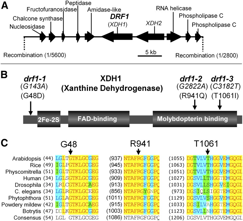
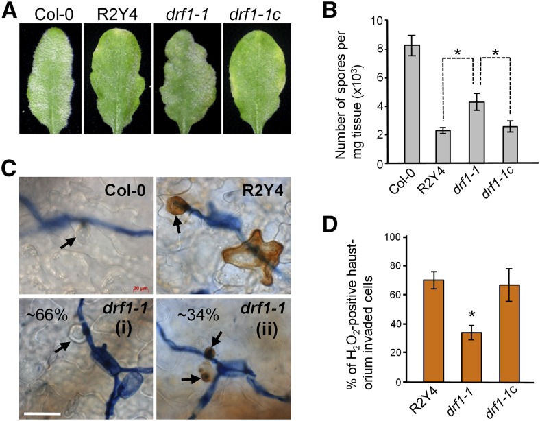
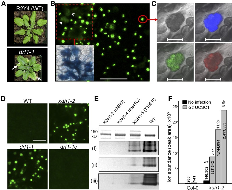
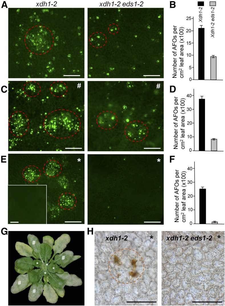
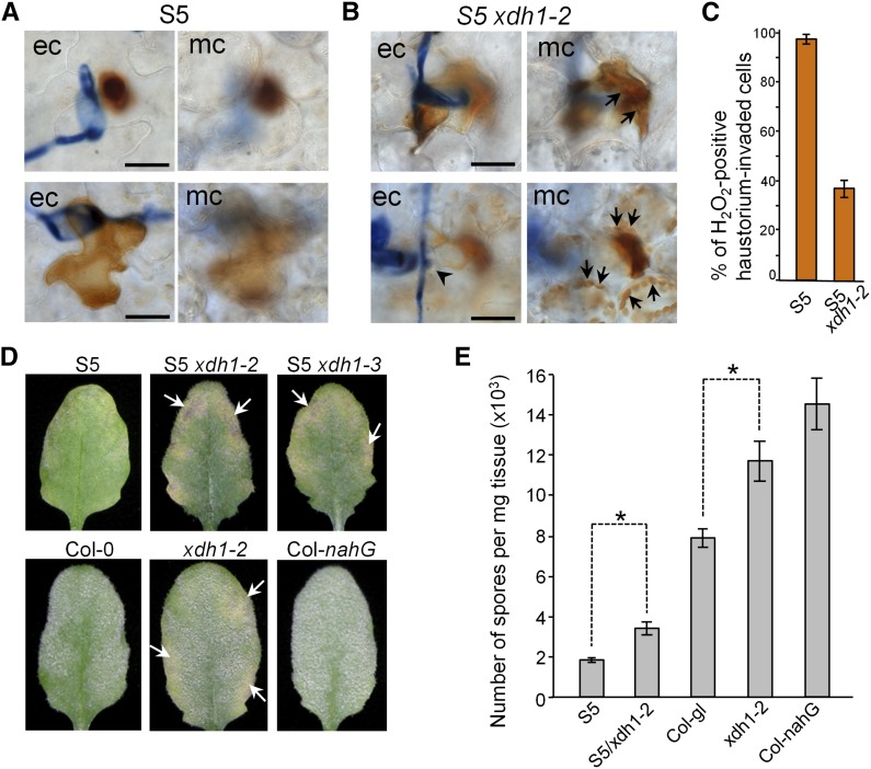
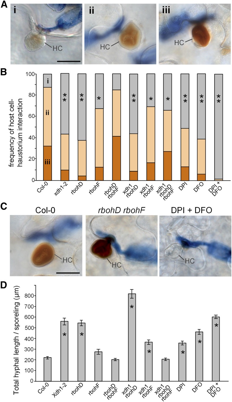
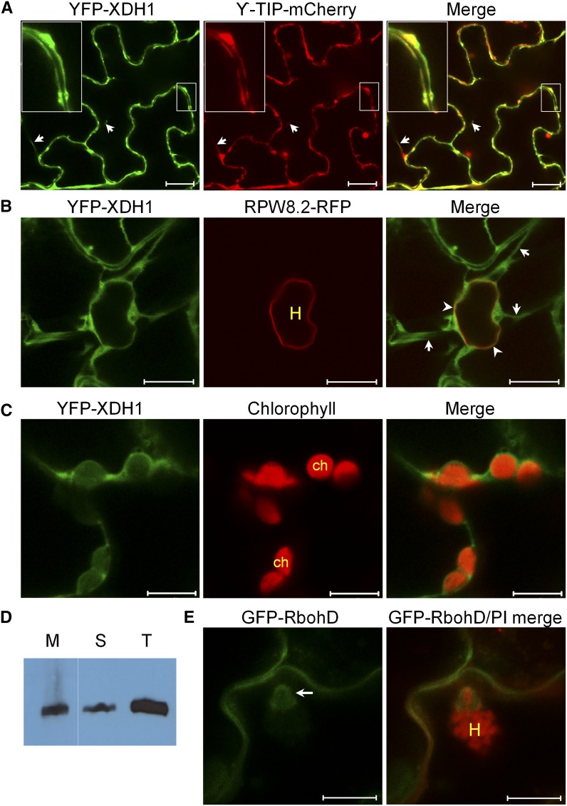
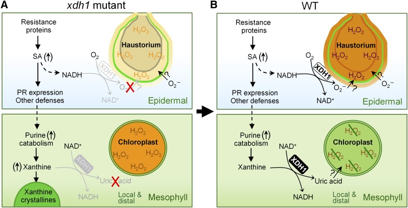
While plants produce reactive oxygen species (ROS) for stress signaling and pathogen defense, they need to remove excessive ROS induced during stress responses in order to minimize oxidative damage. How can plants fine-tune this balance and meet such conflicting needs? Here, we show that XANTHINE DEHYDROGENASE1 (XDH1) in Arabidopsis thaliana appears to play spatially opposite roles to serve this purpose. Through a large-scale genetic screen, we identified three missense mutations in XDH1 that impair XDH1's enzymatic functions and consequently affect the powdery mildew resistance mediated by RESISTANCE TO POWDERY MILDEW8 (RPW8) in epidermal cells and formation of xanthine-enriched autofluorescent objects in mesophyll cells. Further analyses revealed that in leaf epidermal cells, XDH1 likely functions as an oxidase, along with the NADPH oxidases RbohD and RbohF, to generate superoxide, which is dismutated into H2O2 The resulting enrichment of H2O2 in the fungal haustorial complex within infected epidermal cells helps to constrain the haustorium, thereby contributing to RPW8-dependent and RPW8-independent powdery mildew resistance. By contrast, in leaf mesophyll cells, XDH1 carries out xanthine dehydrogenase activity to produce uric acid in local and systemic tissues to scavenge H2O2 from stressed chloroplasts, thereby protecting plants from stress-induced oxidative damage. Thus, XDH1 plays spatially specified dual and opposing roles in modulation of ROS metabolism during defense responses in Arabidopsis.


Figures

Figure 1.
Figure 1.
Cloning of DRF1. (A) Map-based cloning of DRF1 using an F2 population derived from drf1-1 × Ler. (B) The nature and position of the three drf1 mutations in XDH1 that encodes a xanthine dehydrogenase with three functional domains. (C) Protein sequence alignment showing conservation of the three mutated residues (indicated by arrows) among XDH homologs from rice (AAT81740), Physcomitrella patens (EDQ74505), human (NP_000370), Drosophila melanogaster (NP_524337), Caenorhabditis elegans (NP_500531), Phytophthora infestans (EEY63796), powdery mildew (CCU77189), and Botrytis cinerea (CCD52002).
Figure 2.
Figure 2.
Characterization of the Defense Phenotypes of the drf1-1 Mutant. (A) Representative leaves of indicated genotypes showing whitish fungal mass. Six-week-old plants were inoculated with Gc UCSC1 and pictures were taken at 10 dpi. R2Y4 is a homozygous Col-0 line expressing RPW8.2-YFP from the RPW8.2 promoter. drf1-1c is a representative line of drf1-1 genetically complemented with XDH1 expressed from the XDH1 promoter. (B) Quantification of disease susceptibility of plants in (A). Data are means ± se from four replicated experiments. Asterisks indicate significant difference (P < 0.05, n = 4) for the paired comparisons using Tukey’s HSD test following one-way ANOVA. (C) Representative microscopic images of invaded epidermal cells of indicated genotypes showing whole-cell or haustorial complex-confined H2O2 stained by DAB at 52 hpi. Note (i) and (ii) denote two types of reactions of drf1-1 mutant plants. Arrows indicate haustoria. Bar = 50 μm. (D) Frequencies of H2O2-positive epidermal cells (whole-cell H2O2 + haustorium complex-confined H2O2 of indicated genotypes used in [C]). At least 100 haustorium-invaded cells were assessed for each genotype. Data are means ± sd from three replicated experiments. Asterisk indicates significant difference when compared with other two genotypes (P < 0.01; n = 3, Student’s t test).
Figure 3.
Figure 3.
Characterization of AFOs. (A) Compared with the parental R2Y4 line (WT) expressing RPW8.2-YFP, drf1-1 showed slightly reduced stature with shorter petioles and displayed early leaf senescence (arrows). (B) Mature leaves of 7-week-old (or more) drf1-1 plants developed AFOs of various sizes that were subsequently associated with cell death shown by diffuse autofluorescence, indicated by arrowheads and trypan blue staining (inset). Bars = 100 μm. (C) Confocal images of an AFO from (B) showing autofluorescence under 405-, 514-, and 561-nm laser excitation. Bars = 5 μm. (D) Development of AFOs in drf1-1 and an XDH1 knockout line (GK-049D04; xdh1-2) and lack of AFOs in drf1-1 genetically complemented by expressing XDH1 from its native promoter (drf1-1c). Bars = 50 μm. (E) Assay for the enzymatic activities of three mutant proteins, XDH1-3 (G48D), XDH1-4 (R941Q), and XDH1-5 (T1061I), in comparison with XDH1. Recombinant proteins (∼2 μg) purified after heterologous expression in P. pastoris (visualized in gel by Coomassie blue staining in the top panel) were used for measuring (i) the basic XDH activity using hypoxanthine and NAD+ as substrate, (ii) superoxide production using hypoxanthine and O2 as substrate, or (iii) superoxide production using NADH and O2 as substrate (for details, see Methods). (F) Xanthine levels in AFOs from uninfected or Gc UCSC1-infected leaves of 12-week-old xdh1-2 and the equivalent extracts from leaves of 12-week-old Col-0 grown under the same conditions. Leaves were collected at 7 dpi. AFOs were enriched by sucrose gradient centrifugation and subjected to liquid chromatography-mass spectrometry analysis (see Methods). The numbers within or on top of the columns are exact peak areas reporting the total ion abundance. The numbers followed by “x” are fold increase of xanthine levels compared with that indicated by asterisks.
Figure 4.
Figure 4.
EDS1 Is Required for Local and Systemic Amplification of AFO Formation. Eight-week-old plants of xdh1-2 or xdh1-2 eds1-2 were examined for AFO formation. All bars = 100 μm. (A) and (B) AFO formation in mature leaves of uninfected plants from both genotypes as control for (C,D). Note that AFOs in xdh1-2 eds1-2 were in lower density with fewer clusters as seen in xdh1-2 (circled by dashed red lines). (C) and (D) AFO induction by powdery mildew (Gc UCSC1) infection in AFO-free leaves of plants from both genotypes. Images were taken at 14 dpi. (E) and (F) Systemic AFOs in uninfected young leaves (indicated by an asterisk, as seen in [G]) induced by powdery mildew infection on older leaves (indicated by a pound sign, as seen in [G]) of the indicated genotypes from 5 dpi on. No AFOs were found in similar young leaves of uninfected xdh1-2 (inset in [E]). Images were taken at 14 dpi. Data in (B), (D), and (F) represent means ± se, calculated from six similarly aged leaves (two from one plant) for each genotype. (G) One representative 8-week-old plant infected with Gc UCSC1 at 10 dpi. (H) H2O2 accumulation in chloroplasts of uninfected younger leaves (asterisks) revealed by DAB staining.
Figure 5.
Figure 5.
Chloroplast-H2O2 Accumulation in Mesophyll Cells Due to Loss of XDH1 and Its Suppression by Exogenous Uric Acid. (A) Five (1 to 5) serial images showing gradual amplification of chloroplast-H2O2 accumulation revealed by DAB staining in mesophyll cells of mature leaves of 7-week-old xdh1-2 plants and lack of H2O2 accumulation in epidermal cells (5′). Note that initial H2O2 accumulation occurred in one or few chloroplasts (arrows) and H2O2-positive chloroplasts were often disoriented, aggregated, and subsequently degraded (arrowheads). Bars = 50 μm. (B) Three (1 to 3) serial images showing collapse of mesophyll cells whose chloroplasts accumulated H2O2 (inset in 2) 4 to 6 d after AFO formation in mature leaves of xdh1-2 plants stained by trypan blue. Bars = 200 μm. (C) Images showing suppression of chloroplast-H2O2 accumulation in mature leaves of 6-week-old xdh1-2 plants by exogenous uric acid. Fully expanded leaves of xdh1-2 were inserted (with their petioles) into MS-agar medium supplemented with uric acid and cultured for 4 weeks with one transfer per week to fresh MS-agar plates containing the same levels of uric acid. Note that (i) MS-agar conditions, though good for supplying uric acid, attenuated chloroplast-H2O2 production in xdh1-2; and (ii) these images showed the highest local density of H2O2-positive mesophyll cells for each treatment. This experiment was repeated three times with similar results. Bars = 200 μm. (D) Quantification of DAB-positive mesophyll cells in (C). Data are means ± se, calculated from four duplicated leaf samples of one experiment in (C).
Figure 6.
Figure 6.
XDH1 Is Required for RPW8-Mediated and Basal Resistance against Powdery Mildew. (A) and (B) Loss of XDH1 compromised RPW8-dependent H2O2 production in haustorium-invaded epidermal cells (ec) but resulted in chloroplast H2O2 accumulation in underneath mesophyll cells (mc). Leaves from 6-week-old S5 and S5/xdh1-2 were evenly inoculated with Gc UCSC1, subjected to DAB staining at 60 hpi, and imaged with a Zeiss Axio microscope. H2O2 accumulation (reported by brownish DAB staining) was either confined in the haustorial complex or spread throughout the entire invaded epidermal cell (ec) in S5 (A). By contrast, while H2O2 accumulation in haustorium-invaded epidermal cells (arrowhead) was reduced, in ∼10% of penetration sites chloroplast H2O2 accumulation (indicated by arrowhead) occurred in the mesophyll cells underneath the infection site in S5/xdh1-2 at 60 hpi (B). Such induced H2O2 accumulation in affected mesophyll cells increased to >60% after 8 dpi. Bars = 50 µm. (C) Quantitative assessment of H2O2 accumulation in haustorium-invaded epidermal cells. Data represent means ± se of four duplicate experiments in which >200 haustorium-invaded epidermal cells per genotype were examined. (D) Representative leaves from indicated genotypes infected with Gc UCSC1 at 10 dpi. Note chlorotic or necrotic lesions (white arrows) induced by powdery mildew infection in plants impaired for XDH1. (E) Quantification of disease susceptibility of the indicated genotypes. Infected leaves were assayed at 7 dpi before extensive chlorosis occurred in xdh1-2 plants. Data are means ± se, calculated from four duplicate samples per genotype in one of three independent experiments in which similar results were obtained. Asterisks indicate significant difference (P < 0.01; n = 4, Student’s t test).
Figure 7.
Figure 7.
XDH1, RbohD, and RbohF Orchestrate H2O2 Generation in Haustorial Complexes. Single, double, and triple mutants concerning XDH1 and NADPH oxidase-encoding RbohD and RbohF were inoculated with Gc UMSG1, a poorly adapted powdery mildew isolate. H2O2 accumulation in invaded epidermal cells was revealed by DAB staining. (A) Three typical levels of H2O2 accumulation in HCs, i.e., no or very little (i), moderate (ii), and strong (iii) that were observed in most genotypes. Bars = 20 µm. (B) Frequencies of the three levels of HC-H2O2 accumulation in eight indicated genotypes treated with 100 μM DPI and/or 2 mM DFO. Fully expanded leaves of 6-week-old plants were inoculated evenly with Gc UMSG1 and then subjected to DAB staining at 60 hpi to assess HC-H2O2 accumulation. At least 200 HCs were assayed for each genotype. Three different color bars represent the three levels of H2O2 accumulation in (A). Statistical analysis was conducted by comparing all genotypes or treatments to Col-0 wild type using ANOVA in R (www.r-project.org). One asterisk indicates significant difference at P < 0.05; two asterisks indicate significant difference at P < 0.01. This experiment was repeated three times with similar results. (C) Three representative HC-H2O2 of the indicated genotypes. Bars = 20 µm. (D) Basal resistance reflected by the total hyphal length per sporeling in eight indicated genotypes treated with DPI and/or DFO. Data are means ± se, calculated from at least 20 sporelings per genotype in one of three duplicate experiments. The asterisk indicates significant difference when compared with Col-0 wild type (P < 0.01; Student t test).
Figure 8.
Figure 8.
XDH1 Is Membrane Associated and Localized to the Tonoplast. (A) Precise colocalization of YFP-XDH1 with a tonoplast marker γ-TIP-mCherry (Nelson et al., 2007) in leaf epidermal cells of N. benthamiana (transient expression) or Arabidopsis Col-0 (stable expression). YFP-XDH1 was expressed from the 35S promoter and γ-TIP-mCherry was expressed from the maize (Zea mays) ubiquitin promoter. Arrows indicate transvacuolar strands. Bars = 20 µm. (B) Dynamic and intimate association of YFP-XDH1-labeled tonoplast with the EHM. A Col-0 line transgenic for both 35S:YFP-XDH1 and pRPW8.2:RPW8.2-RFP was inoculated with Gc UCSC1. Infected leaves were subjected to confocal imaging at 2 dpi. Note the seemingly partial co-localization between YFP-XDH1 and RPW8.2-RFP at the EHM (arrowheads) and extensive transvacuolar strands (arrows) connecting the distal portion of the tonoplast to the part wrapping the haustorium (H). Bars = 10 µm. (C) Close association of YFP-XDH1-labeled tonoplast with the chloroplasts (ch) visualized by autofluorescence of chlorophyll. Bars = 10 µm. (D) A gel blot assay showing that HA-XDH1 exists in both soluble (S) and membrane (M) fractions. Total protein (T) was extracted from leaves of Arabidopsis plants expressing HA-XDH1 from the 35S promoter, fractionated by ultracentrifugation, gel blotted, and analyzed using an anti-HA antibody. (E) Localization of YFP-RbohD expressed from the native promoter (Hao et al., 2014) to the plasma membrane and possibly the haustorial neck or papilla (arrow). Bars = 10 µm.
Figure 9.
Figure 9.
A Working Model for the Opposing Roles of XDH1 in Epidermal and Mesophyll Cells. Based on our data from xdh1 mutants (A), we propose the following working model (B) to illustrate the dual functions of XDH1. In powdery mildew-infected epidermal cells, following activation of SA-dependent defense signaling (which leads to NADH generation), XDH1 mainly functions as NADH oxidase to produce O2⋅– that is dismutated to H2O2, thereby contributing to H2O2 enrichment in haustorial complexes as part of a basal defense response. By contrast, XDH1 in mesophyll cells carries out its basic enzymatic activity to convert (hypo)xanthine to uric acid. Uric acid is transported via an unknown mechanism to chloroplasts where it protects chloroplasts from stress-induced oxidative damage by scavenging excessive ROS, thereby dampening oxidative bursts in local as well as systemic tissues. Our data also suggest that purine catabolism may be intrinsically geared up (↑) with plant defense responses such that while adequate H2O2 is produced by XDH1 and other oxidases in the epidermis for fighting against pathogen invasion, more uric acid is produced by XDH1 in affected mesophyll cells that scavenge stress-induced H2O2, thereby confining local and systemic defense response.

Comment in

Similar articles

Cited by

References

    1. Aarts N., Metz M., Holub E., Staskawicz B.J., Daniels M.J., Parker J.E. (1998). Different requirements for EDS1 and NDR1 by disease resistance genes define at least two R gene-mediated signaling pathways in Arabidopsis. Proc. Natl. Acad. Sci. USA 95: 10306–10311. - PMC - PubMed
    1. Alvarez M.E., Pennell R.I., Meijer P.J., Ishikawa A., Dixon R.A., Lamb C. (1998). Reactive oxygen intermediates mediate a systemic signal network in the establishment of plant immunity. Cell 92: 773–784. - PubMed
    1. Asada K. (2006). Production and scavenging of reactive oxygen species in chloroplasts and their functions. Plant Physiol. 141: 391–396. - PMC - PubMed
    1. Bartsch M., Gobbato E., Bednarek P., Debey S., Schultze J.L., Bautor J., Parker J.E. (2006). Salicylic acid-independent ENHANCED DISEASE SUSCEPTIBILITY1 signaling in Arabidopsis immunity and cell death is regulated by the monooxygenase FMO1 and the Nudix hydrolase NUDT7. Plant Cell 18: 1038–1051. - PMC - PubMed
    1. Booth V.H. (1935). The identity of xanthine oxidase and the Schardinger enzyme. Biochem. J. 29: 1732–1748. - PMC - PubMed

Publication types

MeSH terms

LinkOut - more resources