

Excited State Electron Distribution and Role of the Terminal Amine in Acidic and Basic Tryptophan Dipeptide Fluorescence
- PMID: 27152052
- PMCID: PMC4852485
- DOI: 10.1016/j.molstruc.2016.03.098
Excited State Electron Distribution and Role of the Terminal Amine in Acidic and Basic Tryptophan Dipeptide Fluorescence
Abstract
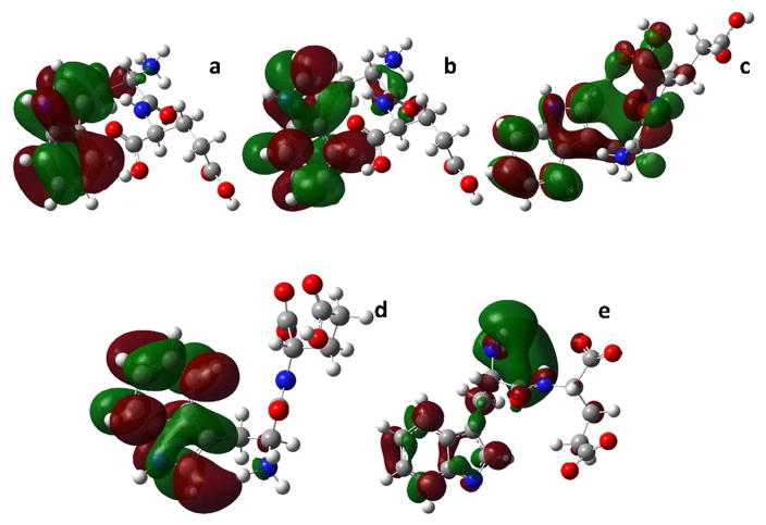
The results of quantum yield (QY) study of tryptophanyl glutamate (Trp-Glu), tryptophanyl lysine (Trp-Lys) and lysinyl tryptophan (Lys-Trp) dipeptides over the pH range, 1.5 - 13, show that the charge state of the N-terminal amine, and not the nominal molecular charge determines the QY. When the terminal amine is protonated, QY is low (10-2) for all three dipeptides. As the terminal amine cation is found proximal to the indole ring in Trp-Glu and Trp-Lys conformers but not in those for Lys-Trp, its effect may lie only in the partitioning of energy between nonradiative processes, not on QY reduction. QY is also low when both the N-terminal amine and indole amine are deprotonated. These two low QY states can be distinguished by fluorescence lifetime measurement. Molecular dynamics simulation shows that the Chi 1 conformers persist for tens of nanoseconds such that 100 - 101 nanosecond lifetimes may be associated with individual Chi 1 conformers. The ground state electron density or isosurface of high QY (0.30) 3-methyindole has a uniform electron density over the indole ring as do the higher QY Trp dipeptide conformers. This validates the association of ground state isosurfaces with QY. Excited state orbitals from calculated high intensity, low energy absorption transitions are typically centered over the indole ring for higher QY dipeptide species and off the ring in lower QY species. Thus excited state orbitals substantiate the earlier finding that the ground state isosurface charge density pattern on the indole ring can be predictive of QY.
Keywords: Tryptophan fluorescence; excited state; fluorescence lifetime; molecular dynamics simulation; quantum mechanics calculations; quantum yield.
Figures


Similar articles
-
The broken ring: reduced aromaticity in Lys-Trp cations and high pH tautomer correlates with lower quantum yield and shorter lifetimes.J Phys Chem B. 2014 Jun 26;118(25):7059-69. doi: 10.1021/jp503355h. Epub 2014 Jun 17. J Phys Chem B. 2014. PMID: 24882092 Free PMC article.
-
Relating Trp-Glu dipeptide fluorescence to molecular conformation: the role of the discrete Chi 1 and Chi 2 angles.J Comput Chem. 2013 Jul 5;34(18):1549-60. doi: 10.1002/jcc.23288. Epub 2013 Apr 8. J Comput Chem. 2013. PMID: 23564660 Free PMC article.
-
Fluorescence of tryptophan in designed hairpin and Trp-cage miniproteins: measurements of fluorescence yields and calculations by quantum mechanical molecular dynamics simulations.J Phys Chem B. 2013 Feb 14;117(6):1790-809. doi: 10.1021/jp3097378. Epub 2013 Feb 4. J Phys Chem B. 2013. PMID: 23330783 Free PMC article.
-
Anomalous temperature fluorescence quenching of N-Trp terminal peptides.Biopolymers. 1995 Dec;36(6):723-33. doi: 10.1002/bip.360360606. Biopolymers. 1995. PMID: 8555420
-
Photoinduced intramolecular tryptophan oxidation and excited-state behavior of [Re(L-AA)(CO)3(α-diimine)](+) (L = pyridine or imidazole, AA = tryptophan, tyrosine, phenylalanine).Inorg Chem. 2011 Jul 4;50(13):6122-34. doi: 10.1021/ic200252z. Epub 2011 Jun 8. Inorg Chem. 2011. PMID: 21650203
References
-
- Burstein E, Vedenkina N, Ivkova M. Fluorescence and the location of tryptophanresidues in protein molecules. Photochem Photobiol. 1973;18:263–279. - PubMed
-
- Callis PR. Exploring the electrostatic landscape of proteins with tryptophan fluorescence. In: Geddes C, editor. Reviews in Fluorescence 2007. Vol. 4. Springer; New York: 2007. pp. 199–248.
-
- Reshetnyak Y, Burstein E. Assignments of the components of the fluorescence spectrum of protein to tryptophan residues based on the properties of their microenvironments in a three dimensional structure. Biophysics. 1997;42:267–274.
-
- Lackowicz J. Principles of fluorescence spectroscopy. Springer Science + Business Media LLC; New York, NY: 2006.
-
- Valeur B. Molecular fluorescence: principles and applications. Wiley-VCH Verlag GmbH; Weinheim, Germany: 2001. p. 339.
Grants and funding
LinkOut - more resources
Full Text Sources
Other Literature Sources
Research Materials