. 2016 Feb:11:94-105.
doi: 10.1016/j.coche.2016.01.012.

Modeling tumor microenvironments using custom-designed biomaterial scaffolds

Affiliations

Modeling tumor microenvironments using custom-designed biomaterial scaffolds

Zen Liu et al. Curr Opin Chem Eng. 2016 Feb.

Abstract

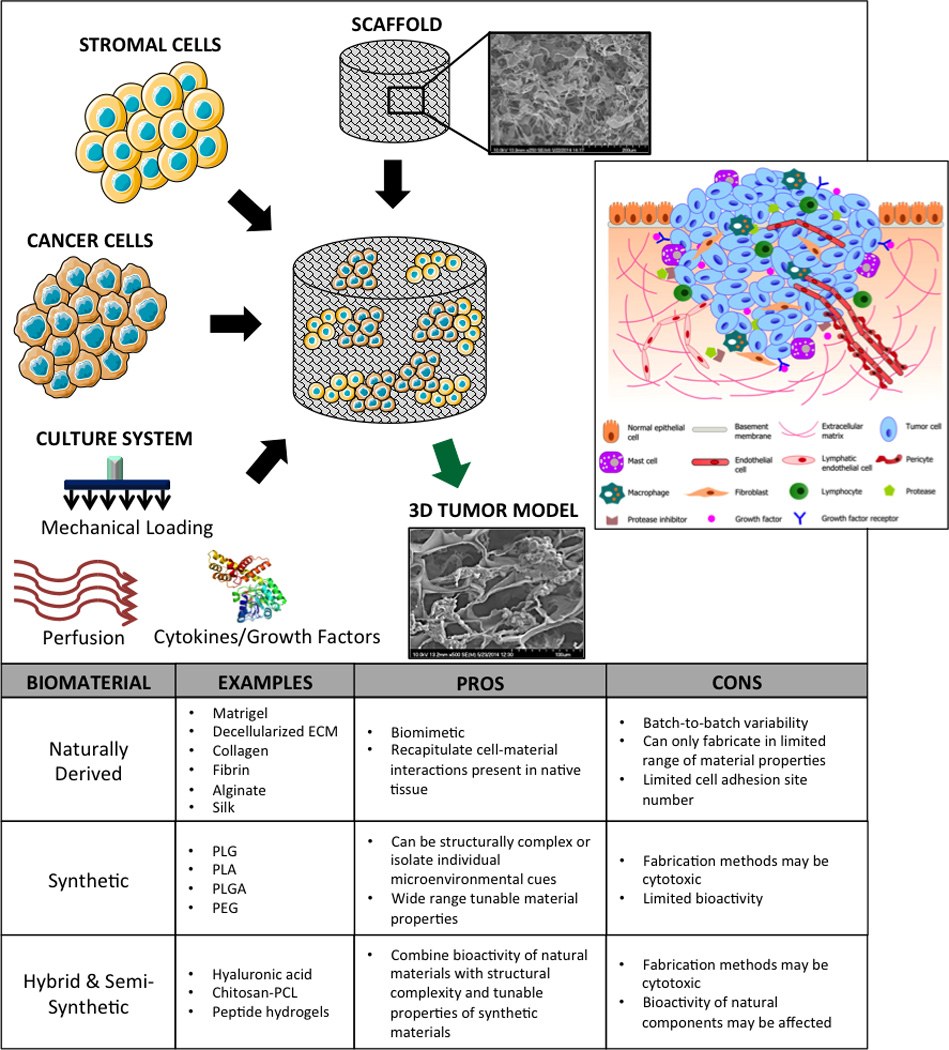
The dominant roles of the tumor microenvironment in regulating tumor formation, progression, and metastasis have driven the application of tissue engineering strategies in cancer biology. Highly dynamic and reciprocal communication of tumor cells with their surroundings suggests that studying cancer in custom-designed biomaterial scaffolds may lead to novel therapeutic targets and therapeutic regimens more reliably than traditional monolayer tissue culture models. As tissue engineering becomes progressively more successful in recapitulating the native tumor environment, critical insights into mechanisms of tumor resistance may be elucidated, to impact clinical practice, drug development, and biological research. We review here the recent developments in the use of custom-designed biomaterial scaffolds for modeling human tumors.

Keywords: Biomaterials; cancer; scaffold; tissue engineering; tumor models.


Figures

Figure 1
Figure 1. Generating a three-dimensional tumor model
Cancer cells, supporting stromal cells, biomaterial scaffold, molecular and physical signals are used in an integrated way to bioengineer in vitro models of human cancer. Inset: illustration of a typical tumor microenvironment with the supporting cells, ECM, and signaling molecules; reproduced with permission from [2].
Figure 2
Figure 2. Naturally derived biomaterials
(a) Digital image of a collagen type I sponge (top) and corresponding scanning electron micrograph (bottom); (b) Digital image of decellularized human adipose tissue sponge (left) and a corresponding scanning electron micrograph (right) [21]; (c) Phase contrast images of tumor spheroids grown in 3D fibrin hydrogels of varying stiffness; reproduced with permission from [31].
Figure 3
Figure 3. Synthetic and semi-synthetic biomaterials
Scanning electron micrographs of (a) porous poly-lactic-glycolic scaffolds; reproduced with permission from [37] and (b) electrospun polycaprolactone scaffold; scale bar = 200 µm; reproduced with permission from [39]. (c) Scanning electron micrograph of a self-assembling peptide hydrogel; reproduced with permission from [49]. (d) Scanning electron micrograph of dehydrated hyaluronic acid gels; scale bar = 20 µm; inset scale bar = 5 µm; reproduced with permission from [45].
Figure 4
Figure 4. Microfabricated 3D substrates
(a, b) Photolithography technique for generating polyacrylamide microchannels; reproduced with permission from [51]. (c) Phase contrast images of U373-MG human glioma cells migrating inside channels of varying stiffness and width, scale bar = 40 µm; reproduced with permission from [51]. (d) 3D microlithography approach to engineering mammary epithelial duct tissue; reproduced with permission from [53]. (e) schematic depicting “duct” versus “end” locations in epithelial host tissue; reproduced with permission from [53].

References

    1. Siegel RL, Miller KD, Jemal A. Cancer statistics, 2015. CA: Cancer J Clin. 2015;65:5–29. - PubMed
    1. Xu X, Farach-Carson MC, Jia X. Three-dimensional in vitro tumor models for cancer research and drug evaluation. Biotechnol Adv. 2014;32:1256–1268. - PMC - PubMed
    1. Bissell MJ, Hines WC. Why don’t we get more cancer? A proposed role of the microenvironment in restraining cancer progression. Nat Med. 2011;17:320–329. - PMC - PubMed
    1. Levental KR, Yu H, Kass L, Lakins JN, Egeblad M, Erler JT, Fong SF, Csiszar K, Giaccia A, Weaver VM. Matrix crosslinking forces tumor progression by enhancing integrin signaling. Cell. 2009;139:891–906. - PMC - PubMed
    1. Provenzano PP, Eliceiri KW, Campbell JM, Inman DR, White JG, Keely PJ. Collagen reorganization at the tumor-stromal interface facilitates local invasion. BMC Med. 2006;4:38–54. - PMC - PubMed