. 2016 May 6;11(5):e0154427.
doi: 10.1371/journal.pone.0154427. eCollection 2016.

Melatonin Preserves Blood-Brain Barrier Integrity and Permeability via Matrix Metalloproteinase-9 Inhibition

Affiliations

Melatonin Preserves Blood-Brain Barrier Integrity and Permeability via Matrix Metalloproteinase-9 Inhibition

Himakarnika Alluri et al. PLoS One. .

Abstract

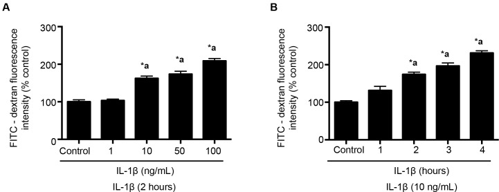
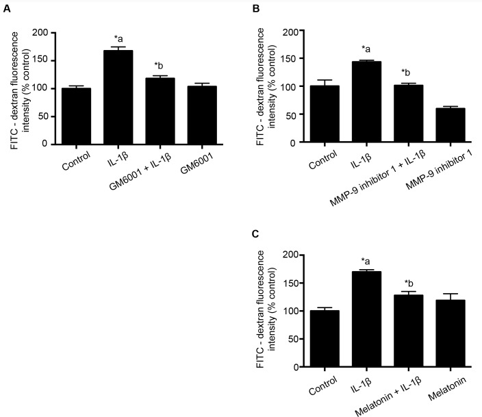
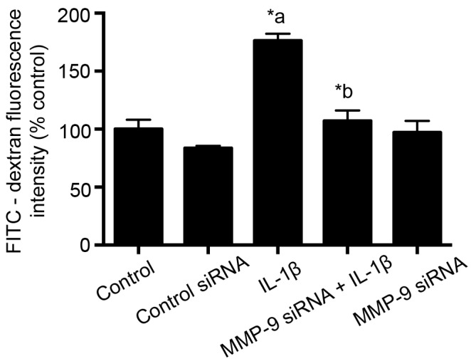
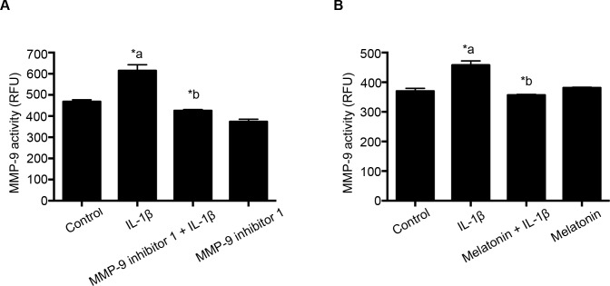
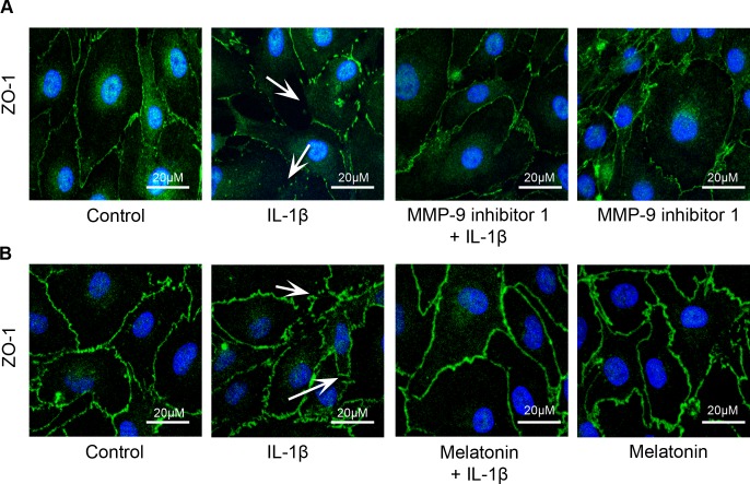
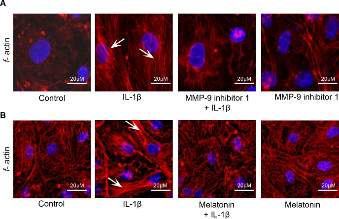
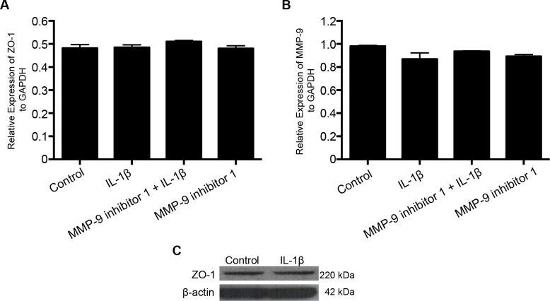
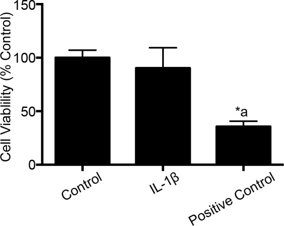
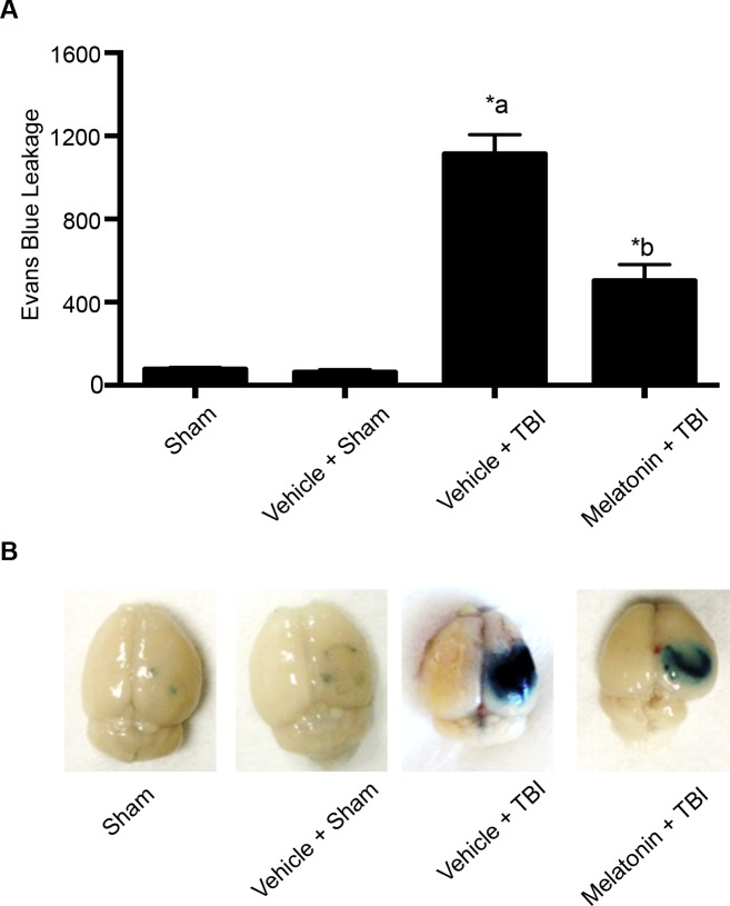
Microvascular hyperpermeability that occurs at the level of the blood-brain barrier (BBB) often leads to vasogenic brain edema and elevated intracranial pressure following traumatic brain injury (TBI). At a cellular level, tight junction proteins (TJPs) between neighboring endothelial cells maintain the integrity of the BBB via TJ associated proteins particularly, zonula occludens-1 (ZO-1) that binds to the transmembrane TJPs and actin cytoskeleton intracellularly. The pro-inflammatory cytokine, interleukin-1β (IL-1β) as well as the proteolytic enzymes, matrix metalloproteinase-9 (MMP-9) are key mediators of trauma-associated brain edema. Recent studies indicate that melatonin a pineal hormone directly binds to MMP-9 and also might act as its endogenous inhibitor. We hypothesized that melatonin treatment will provide protection against TBI-induced BBB hyperpermeability via MMP-9 inhibition. Rat brain microvascular endothelial cells grown as monolayers were used as an in vitro model of the BBB and a mouse model of TBI using a controlled cortical impactor was used for all in vivo studies. IL-1β (10 ng/mL; 2 hours)-induced endothelial monolayer hyperpermeability was significantly attenuated by melatonin (10 μg/mL; 1 hour), GM6001 (broad spectrum MMP inhibitor; 10 μM; 1 hour), MMP-9 inhibitor-1 (MMP-9 specific inhibitor; 5 nM; 1 hour) or MMP-9 siRNA transfection (48 hours) in vitro. Melatonin and MMP-9 inhibitor-1 pretreatment attenuated IL-1β-induced MMP-9 activity, loss of ZO-1 junctional integrity and f-actin stress fiber formation. IL-1β treatment neither affected ZO-1 protein or mRNA expression or cell viability. Acute melatonin treatment attenuated BBB hyperpermeability in a mouse controlled cortical impact model of TBI in vivo. In conclusion, one of the protective effects of melatonin against BBB hyperpermeability occurs due to enhanced BBB integrity via MMP-9 inhibition. In addition, acute melatonin treatment provides protection against BBB hyperpermeability in a mouse model of TBI indicating its potential as a therapeutic agent for brain edema when established in humans.


Conflict of interest statement

Competing Interests: The authors have declared that no competing interests exist.

Figures

Fig 1
Fig 1. IL-1β treatment induces dose and time dependent increase in monolayer hyperpermeability.
In Panel A, IL-1β treatment at doses 10, 50 and 100 ng/mL for 2 hours are shown to significantly increase BBB permeability compared to the control group (n = 4; p<0.05). Panel B indicates significant increase in IL-1β induced BBB permeability at 2, 3 and 4 hours compared to the control (n = 4; p<0.05). Monolayer permeability is expressed as a percentage control of FITC-dextran-10 kDa fluorescent intensity, plotted on the Y-axis. Data are expressed as mean ± % SEM. ‘*a’ indicates significant increase compared to the control group.
Fig 2
Fig 2. GM6001, MMP-9 inhibitor 1 and melatonin pretreatment attenuates IL-1β treatment-induced monolayer hyperpermeability.
Panel A indicates the effect of GM6001 (broad-spectrum MMP inhibitor; n = 4); while Panels B and C employ MMP-9 specific inhibitors: MMP-9 inhibitor 1 (n = 4) and melatonin (n = 6) pretreatment on IL-1β (10 ng/mL; 2 hours)—induced monolayer hyperpermeability. Monolayer permeability is expressed as a percentage control of FITC-dextran-10 kDa fluorescence intensity, plotted on the Y-axis. Data are expressed as mean ± % SEM. ‘*a’ indicates significant increase compared to control group; ‘*b’ indicates significant decrease compared to the IL-1β treated group. p<0.05 was considered statistically significant.
Fig 3
Fig 3. Knockdown of MMP-9 by siRNA attenuates IL-1β treatment-induced monolayer hyperpermeability.
Monolayer permeability is expressed as percentage flux of FITC-dextran-10 kDa fluorescence intensity, plotted on the Y-axis. Data are expressed as mean ± % SEM. ‘*a’ indicates significant increase compared to the control group; ‘*b’ indicates significant decrease compared to the IL-1β (10 ng/mL; 2 hours) treatment group. siRNA transfected groups were compared to control siRNA transfected group (n = 4; p<0.05).
Fig 4
Fig 4. MMP-9 inhibitor 1 and melatonin pretreatment attenuates IL-1β treatment- induced MMP-9 activity.
MMP-9 inhibitor 1 (n = 4) and melatonin (n = 5) pretreatment attenuated IL-1β treatment-induced MMP-9 activity in RBMECs. MMP-9 activity is expressed as relative fluorescence units (RFU), plotted on the Y-axis. Data are expressed as mean ± SEM. ‘*a’ indicates significant increase compared to the control group; ‘*b’ indicates significant decrease compared to the IL-1β (10 ng/mL; 2 hours) treatment group. p<0.05 was considered statistically significant.
Fig 5
Fig 5. MMP-9 inhibitor 1 and melatonin pretreatment protects against IL-1β treatment-induced loss of ZO-1 junctional integrity.
IL-1β (10 ng/mL; 2 hours) treatment-induced ZO-1 junctional disruption (white arrows) was decreased on pretreatment with MMP-9 inhibitor 1 (n = 4) and melatonin (n = 4).
Fig 6
Fig 6. MMP-9 inhibitor 1 and melatonin pretreatment reduces IL-1β treatment- induced f-actin stress fiber formation.
IL-1β (10 ng/mL; 2 hours) treatment-induced f-actin stress fiber formation (white arrows) was decreased by pretreatment with MMP-9 inhibitor 1 (n = 4) and melatonin (n = 4).
Fig 7
Fig 7. IL-1β treatment does not induce ZO-1 mRNA or protein expression.
IL-1β (10 ng/mL; 2 hours) treatment neither induces ZO-1/MMP-9 mRNA expression (n = 3) nor alter ZO-1 protein expression (n = 4). RT-PCR data plotted on the Y-axis are expressed as relative expression of ZO-1 normalized to GAPDH. Data are represented as mean ± SEM.
Fig 8
Fig 8. IL-1β treatment does not induce cell death.
IL-1β (10 ng/mL; 2 hours) treatment had no effect on cell viability (n = 5). Hydrogen peroxide (used as a positive control) treatment decreases cell viability significantly (p<0.05). Data are expressed as mean ± % SEM. ‘*’ indicates statistical significance. ‘*a’ indicates significant decrease compared to the control group.
Fig 9
Fig 9
Melatonin pretreatment attenuates TBI-induced BBB hyperpermeability studied by Evans blue dye extravasation method (Panel A). Pictorial representation of the brain tissue from various groups is shown in Panel 9B. Sham injury group was used as the baseline for all comparisons. Melatonin (10 μg/gram body weight of the animal) pretreatment significantly attenuated TBI-induced Evans blue leakage into the extravascular tissue space (p<0.05). Animals were divided into sham (n = 6), vehicle + sham (n = 6), vehicle + TBI (n = 5) and melatonin + TBI (n = 6). Data are expressed as ng/brain cortex ± SEM. ‘*’ indicates statistical significance. ‘a’ indicates significant increase compared to the sham injury/vehicle + sham injury group and ‘b’ indicates significant decrease compared to the vehicle + TBI group.

References

    1. Alluri H, Wiggins-Dohlvik K, Davis ML, Huang JH, Tharakan B (2015) Blood-brain barrier dysfunction following traumatic brain injury. Metab Brain Dis 30: 1093–1104. 10.1007/s11011-015-9651-7 - DOI - PubMed
    1. Bolton SJ, Anthony DC, Perry VH (1998) Loss of the tight junction proteins occludin and zonula occludens-1 from cerebral vascular endothelium during neutrophil-induced blood-brain barrier breakdown in vivo. Neuroscience 86: 1245–1257. - PubMed
    1. Rigor RR, Beard RS Jr., Litovka OP, Yuan SY (2012) Interleukin-1beta-induced barrier dysfunction is signaled through PKC-theta in human brain microvascular endothelium. Am J Physiol Cell Physiol 302: C1513–1522. 10.1152/ajpcell.00371.2011 - DOI - PMC - PubMed
    1. Simi A, Tsakiri N, Wang P, Rothwell NJ (2007) Interleukin-1 and inflammatory neurodegeneration. Biochem Soc Trans 35: 1122–1126. - PubMed
    1. Didier N, Romero IA, Créminon C, Wijkhuisen A, Grassi J, Mabondzo A. (2004) Secretion of interleukin-1β by astrocytes mediates endothelin-1 and tumour necrosis factor-α effects on human brain microvascular endothelial cell permeability. Journal of Neurochemistry 86: 246–254. - PubMed

Publication types

LinkOut - more resources