Tumor-specific cell-cycle decoy by Salmonella typhimurium A1-R combined with tumor-selective cell-cycle trap by methioninase overcome tumor intrinsic chemoresistance as visualized by FUCCI imaging
- PMID: 27152859
- PMCID: PMC4957602
- DOI: 10.1080/15384101.2016.1181240
Tumor-specific cell-cycle decoy by Salmonella typhimurium A1-R combined with tumor-selective cell-cycle trap by methioninase overcome tumor intrinsic chemoresistance as visualized by FUCCI imaging
Abstract
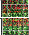
We previously reported real-time monitoring of cell cycle dynamics of cancer cells throughout a live tumor intravitally using a fluorescence ubiquitination cell cycle indicator (FUCCI). Approximately 90% of cancer cells in the center and 80% of total cells of an established tumor are in G0/G1 phase. Longitudinal real-time FUCCI imaging demonstrated that cytotoxic agents killed only proliferating cancer cells at the surface and, in contrast, and had little effect on the quiescent cancer cells. Resistant quiescent cancer cells restarted cycling after the cessation of chemotherapy. Thus cytotoxic chemotherapy which targets cells in S/G2/M, is mostly ineffective on solid tumors, but causes toxic side effects on tissues with high fractions of cycling cells, such as hair follicles, bone marrow and the intestinal lining. We have termed this phenomenon tumor intrinsic chemoresistance (TIC). We previously demonstrated that tumor-targeting Salmonella typhimurium A1-R (S. typhimurium A1-R) decoyed quiescent cancer cells in tumors to cycle from G0/G1 to S/G2/M demonstrated by FUCCI imaging. We have also previously shown that when cancer cells were treated with recombinant methioninase (rMETase), the cancer cells were selectively trapped in S/G2, shown by cell sorting as well as by FUCCI. In the present study, we show that sequential treatment of FUCCI-expressing stomach cancer MKN45 in vivo with S. typhimurium A1-R to decoy quiescent cancer cells to cycle, with subsequent rMETase to selectively trap the decoyed cancer cells in S/G2 phase, followed by cisplatinum (CDDP) or paclitaxel (PTX) chemotherapy to kill the decoyed and trapped cancer cells completely prevented or regressed tumor growth. These results demonstrate the effectiveness of the praradigm of "decoy, trap and shoot" chemotherapy.
Keywords: FUCCI; Salmonella typhimurium A1-R; cancer; cell-cycle; cisplatinum; decoy; methioninase; nude mice; paclitaxel; stomach cancer; trap.
Figures


Similar articles
-
FUCCI Real-Time Cell-Cycle Imaging as a Guide for Designing Improved Cancer Therapy: A Review of Innovative Strategies to Target Quiescent Chemo-Resistant Cancer Cells.Cancers (Basel). 2020 Sep 17;12(9):2655. doi: 10.3390/cancers12092655. Cancers (Basel). 2020. PMID: 32957652 Free PMC article. Review.
-
Methioninase Cell-Cycle Trap Cancer Chemotherapy.Methods Mol Biol. 2019;1866:133-148. doi: 10.1007/978-1-4939-8796-2_11. Methods Mol Biol. 2019. PMID: 30725413
-
Tumor-targeting Salmonella typhimurium A1-R combined with recombinant methioninase and cisplatinum eradicates an osteosarcoma cisplatinum-resistant lung metastasis in a patient-derived orthotopic xenograft (PDOX) mouse model: decoy, trap and kill chemotherapy moves toward the clinic.Cell Cycle. 2018;17(6):801-809. doi: 10.1080/15384101.2018.1431596. Epub 2018 Apr 10. Cell Cycle. 2018. PMID: 29374999 Free PMC article.
-
Tumor-targeting Salmonella typhimurium A1-R decoys quiescent cancer cells to cycle as visualized by FUCCI imaging and become sensitive to chemotherapy.Cell Cycle. 2014;13(24):3958-63. doi: 10.4161/15384101.2014.964115. Cell Cycle. 2014. PMID: 25483077 Free PMC article.
-
Development of recombinant methioninase to target the general cancer-specific metabolic defect of methionine dependence: a 40-year odyssey.Expert Opin Biol Ther. 2015 Jan;15(1):21-31. doi: 10.1517/14712598.2015.963050. Epub 2014 Dec 2. Expert Opin Biol Ther. 2015. PMID: 25439528 Review.
Cited by
-
The Combination of Tumor-targeting Salmonella typhimurium A1-R Plus the Autophagy-inhibitor Chloroquine Synergistically Eradicates HT1080 Fibrosarcoma Cells In Vitro and In Vivo.In Vivo. 2025 Jan-Feb;39(1):102-109. doi: 10.21873/invivo.13807. In Vivo. 2025. PMID: 39740880 Free PMC article.
-
FUCCI Real-Time Cell-Cycle Imaging as a Guide for Designing Improved Cancer Therapy: A Review of Innovative Strategies to Target Quiescent Chemo-Resistant Cancer Cells.Cancers (Basel). 2020 Sep 17;12(9):2655. doi: 10.3390/cancers12092655. Cancers (Basel). 2020. PMID: 32957652 Free PMC article. Review.
-
Anti-tumor activity of an immunotoxin (TGFα-PE38) delivered by attenuated Salmonella typhimurium.Oncotarget. 2017 Jun 6;8(23):37550-37560. doi: 10.18632/oncotarget.17197. Oncotarget. 2017. PMID: 28473665 Free PMC article.
-
Accurate and Safe Tumor Targeting of Orally-administered Salmonella typhimurium A1-R Leads to Regression of an Aggressive Fibrosarcoma in Nude Mice.In Vivo. 2024 Nov-Dec;38(6):2601-2609. doi: 10.21873/invivo.13736. In Vivo. 2024. PMID: 39477429 Free PMC article.
-
Perspectives on Oncolytic Salmonella in Cancer Immunotherapy-A Promising Strategy.Front Immunol. 2021 Feb 25;12:615930. doi: 10.3389/fimmu.2021.615930. eCollection 2021. Front Immunol. 2021. PMID: 33717106 Free PMC article. Review.
References
-
- Yano S, Zhang Y, Miwa S, Tome Y, Hiroshima Y, Uehara F, Yamamoto M, Suetsugu A, Kishimoto H, Tazawa H, et al.. Spatial-temporal FUCCI imaging of each cell in a tumor demonstrates locational dependence of cell cycle dynamics and chemoresponsiveness. Cell Cycle 2014; 13:2110-9; PMID:24811200; http://dx.doi.org/10.4161/cc.29156 - DOI - PMC - PubMed
-
- Sakaue-Sawano A, Kurokawa H, Morimura T, Hanyu A, Hama H, Osawa H, Kashiwagi S, Fukami K, Miyata T, Miyoshi H, et al.. Visualizing spatiotemporal dynamics of multicellular cell cycle progression. Cell 2008; 132:487-98; PMID:18267078; http://dx.doi.org/10.1016/j.cell.2007.12.033 - DOI - PubMed
-
- Zhang Y, Zhang N, Zhao M, Hoffman RM. Comparison of the selective targeting efficacy of Salmonella typhimurium A1-R and VNP20009 on the Lewis lung carcinoma in nude mice. Oncotarget 2015; 6:14625-31; PMID:25714030; http://dx.doi.org/10.18632/oncotarget.3342 - DOI - PMC - PubMed
-
- Hoffman RM, editor. Bacterial Therapy of Cancer: Methods and Protocols Methods in Molecular Biology 1409. Walker John M, series ed. Humana Press; (Springer Science + Business Media; New York: ), 2016
-
- Zhao M, Yang M, Li XM, Jiang P, Baranov E, Li S, Xu M, Penman S, Hoffman RM. Tumor-targeting bacterial therapy with amino acid auxotrophs of GFP-expressing Salmonella typhimurium. Proc Natl Acad Sci USA 2005; 102:755-60; PMID:15644448; http://dx.doi.org/10.1073/pnas.0408422102 - DOI - PMC - PubMed
Publication types
MeSH terms
Substances
LinkOut - more resources
Full Text Sources
Other Literature Sources
