Review
. 2016 May 3;8(5):134.
doi: 10.3390/toxins8050134.

Clostridium difficile Toxins A and B: Insights into Pathogenic Properties and Extraintestinal Effects

Affiliations
Review

Clostridium difficile Toxins A and B: Insights into Pathogenic Properties and Extraintestinal Effects

Stefano Di Bella et al. Toxins (Basel). .

Abstract

Clostridium difficile infection (CDI) has significant clinical impact especially on the elderly and/or immunocompromised patients. The pathogenicity of Clostridium difficile is mainly mediated by two exotoxins: toxin A (TcdA) and toxin B (TcdB). These toxins primarily disrupt the cytoskeletal structure and the tight junctions of target cells causing cell rounding and ultimately cell death. Detectable C. difficile toxemia is strongly associated with fulminant disease. However, besides the well-known intestinal damage, recent animal and in vitro studies have suggested a more far-reaching role for these toxins activity including cardiac, renal, and neurologic impairment. The creation of C. difficile strains with mutations in the genes encoding toxin A and B indicate that toxin B plays a major role in overall CDI pathogenesis. Novel insights, such as the role of a regulator protein (TcdE) on toxin production and binding interactions between albumin and C. difficile toxins, have recently been discovered and will be described. Our review focuses on the toxin-mediated pathogenic processes of CDI with an emphasis on recent studies.

Keywords: Clostridium difficile; pathogenesis; toxins.


Figures
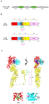
Figure 1
Figure 1
(A) Schematic representation of the PaLoc region containing the following genes: tcdR, tcdB, tcdE, tcdC, and tcdA. Below each gene, the number of base pairs (bp) is indicated. (B) Schematic representation of the multi-modular domain structure described as the ABCD model (A: biological activity; B: binding; C: cutting; D: delivery) domain structure of TcdA and TcdB. The two toxins are composed of: the A domain, corresponding to the N-terminal glucosyltransferase domain (GTD) (red) [32,33]; the C domain, corresponding to the cysteine protease domain (CPD) (cyan) and the three-helix bundle domain (3HB) (blue) identified in TcdA [34], and possibly present also in TcdB; the D domain, which corresponds to the delivery hydrophobic domain (DD) (yellow), containing the small globular domain (SGD) in TcdA (green and yellow diagonal lines) [34] that corresponds to the minimal pore forming region (MPFR) in TcdB (green and yellow diagonal lines) [35], and overlapping only partially (purple and green diagonal lines) to the hydrophobic region (HR) of TcdA [36] and TcdB [35] (purple and yellow diagonal lines); the B domain, corresponding to the receptor binding domain (RBD) (pink) [34,37]. (C) Top: two three-dimensional structures of TcdA are shown, rotated 90° each other (red: GTD; cyan: CPD; blue: 3HB; yellow: DD; pink: RBD), with a detail of the N-terminal region of the D domain showing the SGD and HR; right bottom: detail of the loop containing the amino acid residues 516–522 of the GTD, in which the conserved Ser517 residue forms a hydrogen bond with the phosphate group present in the UDP-glucose, and the conserved Trp519 forms a hydrogen bond with the glycosidic oxygen (PDB: 3SRZ; [18]); left bottom: detail of the catalytic triad Asp589-Hys655-Cys700 of the CPD, which is responsible for the proteolytic cleavage of the toxin dependent upon inositol hexakisphosphate (Insp6) (purple) binding (PDB: 3HO6; [38]). The three-dimensional structures were drawn with the UCSF-Chimera package [39]. For details, see text.
Figure 2
Figure 2
Toxins delivery into the host cell cytosol can be divided into seven main steps: (1) toxin binding to the host cell surface receptor; (2) toxins internalization through a receptor-mediated endocytosis; (3) endosome acidification; (4) pore formation; (5) GTD release from the endosome to the host cell cytoplasm; (6) Rho GTPases inactivation by glucosylation; and (7) downstream effects within the host cell, i.e., toxins-induced cytopathic and cytotoxic effects. For clarity, the color codes used to depict the diverse toxins domains are the same used in Figure 1: GTD: N-terminal glucosyltransferase domain (red); CPD: cysteine protease domain (cyan); DD: delivery domain (yellow).

References

    1. Magill S.S., Edwards J.R., Bamberg W., Beldavs Z.G., Dumyati G., Kainer M.A., Lynfield R., Maloney M., McAllister-Hollod L., Nadle J., et al. Multistate point-prevalence survey of health care-associated infections. N. Engl. J. Med. 2014;370:1198–1208. doi: 10.1056/NEJMoa1306801. - DOI - PMC - PubMed
    1. Bartlett J.G., Gerding D.N. Clinical recognition and diagnosis of Clostridium difficile infection. Clin. Infect. Dis. 2008;46:S12–S18. doi: 10.1086/521863. - DOI - PubMed
    1. Redelings M.D., Sorvillo F., Mascola L. Increase in Clostridium difficile-related mortality rates, United States, 1999–2004. Emerg. Infect. Dis. 2007;13:1417–1419. doi: 10.3201/eid1309.061116. - DOI - PMC - PubMed
    1. Burns D.A., Heap J.T., Minton N.P. Clostridium difficile spore germination: An update. Res. Microbiol. 2010;161:730–734. doi: 10.1016/j.resmic.2010.09.007. - DOI - PubMed
    1. Anthony D.M., Reynolds T., Paton J., Rafter L. Serum albumin in risk assessment for Clostridium difficile. J. Hosp. Infect. 2009;71:378–379. doi: 10.1016/j.jhin.2008.12.003. - DOI - PubMed