. 2016 May 5;98(5):1020-1029.
doi: 10.1016/j.ajhg.2016.03.013.

Mutations in DNMT3B Modify Epigenetic Repression of the D4Z4 Repeat and the Penetrance of Facioscapulohumeral Dystrophy

Affiliations

Mutations in DNMT3B Modify Epigenetic Repression of the D4Z4 Repeat and the Penetrance of Facioscapulohumeral Dystrophy

Marlinde L van den Boogaard et al. Am J Hum Genet. .

Abstract

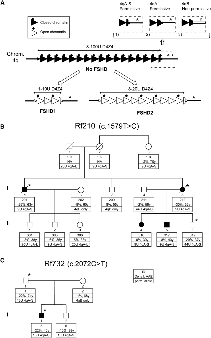
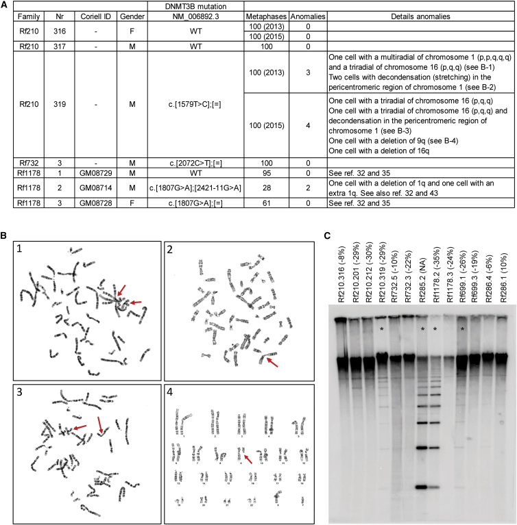
Facioscapulohumeral dystrophy (FSHD) is associated with somatic chromatin relaxation of the D4Z4 repeat array and derepression of the D4Z4-encoded DUX4 retrogene coding for a germline transcription factor. Somatic DUX4 derepression is caused either by a 1-10 unit repeat-array contraction (FSHD1) or by mutations in SMCHD1, which encodes a chromatin repressor that binds to D4Z4 (FSHD2). Here, we show that heterozygous mutations in DNA methyltransferase 3B (DNMT3B) are a likely cause of D4Z4 derepression associated with low levels of DUX4 expression from the D4Z4 repeat and increased penetrance of FSHD. Recessive mutations in DNMT3B were previously shown to cause immunodeficiency, centromeric instability, and facial anomalies (ICF) syndrome. This study suggests that transcription of DUX4 in somatic cells is modified by variations in its epigenetic state and provides a basis for understanding the reduced penetrance of FSHD within families.


Figures

Figure 1
Figure 1
D4Z4 Locus and FSHD2-Affected Families (A) Schematic representation of the D4Z4 locus. In control individuals, the D4Z4 repeat array ranges from 8 to 100 units and shows characteristics of a closed chromatin structure (black triangles) characterized by high CpG methylation, among other things. For both FSHD1 and FSHD2, the chromatin adopts a more open configuration (white triangles) marked by a loss of CpG methylation and other chromatin changes. FSHD1 is caused by a contraction of the D4Z4 repeat to 1–10 units, whereas FSHD2 involves chromatin relaxation due to mutations that affect a chromatin modifier (black dots), most often SMCHD1. The chromatin relaxation must occur in a permissive 4qA (marked by 4qA-S in this figure) or 4qA-L chromosomal region to cause FSHD, given that 4qB chromosomes are non-permissive for FSHD (chromosome 4 variants are displayed in the dashed boxes). 4qA-S and 4qA-L differ by the length of the last partial D4Z4 unit, and protein studies have demonstrated production of DUX4 from both 4qA variants. The 3′ UTR of DUX4 is missing in 4qB chromosomal regions (white square in dashed box), which makes them non-permissive to DUX4 expression. (B and C) Pedigrees of families Rf210 (B) and Rf732 (C). Clinically affected individuals are indicated in black. The key shows the family identifier (ID), Delta1 score, age at examination (AAE), and size of the smallest D4Z4 repeat array on a FSHD-permissive allele (4qA-S and 4qA-L). Additionally, it indicates when no permissive allele was present (4qB only). The cDNA position behind the family ID indicates the cDNA position of the DNMT3B mutation (GenBank: NM_006892.3) present in this family. The asterisk indicates individuals carrying the DNMT3B mutation.
Figure 2
Figure 2
DNMT3B Mutations in FSHD2 (A) Schematic representation of DNMT3B. The amino acid changes (GenBank: NP_008823.1) found in FSHD2-affected families are indicated in red. (B and C) Sanger sequence confirmation of DNMT3B variants (GenBank: NM_006892.3) in Rf210 and Rf732. (D and E) Multiple-sequence alignment (MSA) of DNMT3B across distinct species for DNMT3B variants in Rf210 and Rf732. MSA was performed with ClustalOmega, and alignment was viewed in Jalview and colored as in ClustalX. (F) Ribbon representation of the nuclear-magnetic-resonance structure of the ADD domain of ATRX (PDB: 2JM1). The cysteine residues are shown as sticks. Cys527 is shown in magenta. Zinc ions are represented as spheres. (G) Ribbon representation of the crystallography structure of the C-terminal domain of DNMT3A (chain A [PDB: 2QRV]). The proline residues are shown as sticks. Pro691 is shown in magenta.
Figure 3
Figure 3
Pedigrees of Families Rf286, Rf699, Rf1178, Rf285, and Rf614, Affected by Autosomal-Recessive ICF1 Affected individuals are indicated in black, and DNMT3B mutations (GenBank: NM_006892.3) are shown below each individual. Their clinical phenotypes and DNMT3B mutations have been described before., , , , The key description is identical to that in Figure 1.
Figure 4
Figure 4
DUX4 Presence in FSHD and ICF1 (A) Expression of MYOG, MYH3, DUX4, and LEUTX (DUX4 target) by qPCR in GFP (G)- or MyoD (M)-lentivirus-transduced fibroblasts from control individuals, FSHD1 and FSHD2 cell lines, and individuals Rf210.319, Rf732.3, and ICF-affected Rf1178.2. All transductions were performed twice for each cell line, except for control individual 4 (1× transduced with GFP and 2× transduced with MyoD) and FSHD2-2 (transduced 1× with GFP and 1× with MyoD). Mean expression values with SDs are shown in relation to those of the reference genes GUSB and RPL27. DUX4 was measured with primers for the most common DUX4-4A-S variant, but the primers did not recognize DUX4-4A-L. The fibroblasts from control individual 4 and Rf1178.2 carry a 4qA-L allele and were therefore excluded from analysis of DUX4 expression. Primers are listed in Table S5. (B) Immunofluorescent staining for DUX4 and Myosin in fixed ICF1 myotubes from Rf285.1 (Figure 3) shows DUX4 immunoreactivity in a small percentage of myotubes.
Figure 5
Figure 5
Metaphase Analysis and NBL2 Southern Blot Analysis of Rf210, Rf732, and ICF1-Affected Families (A) Metaphases were analyzed from three heterozygous DNMT3B-mutation carriers (Rf210.319, Rf732.3, and Rf1178.3), one ICF1 individual (Rf1178.2), and three individuals without a DNMT3B variant (Rf210.316, Rf210.317, and Rf1178.1). Identifiers from Leiden University Medical Center and Coriell, the mutation in DNMT3B (GenBank: NM_006892.3), and the number of analyzed metaphases are indicated. Chromosomal anomalies are listed in the last column. (B) Four panels show examples of chromosomal anomalies identified in individual Rf210.319. Chromosomal anomalies are indicated with red arrows. (C) NBL2 Southern blot analysis in Rf210, Rf732, and ICF1-affected families after digestion of 2 μg genomic DNA with the methylation-sensitive endonuclease Eco52I according to previously described protocols. Numbers correspond with pedigrees in Figures 1 and 3. Delta1 scores are indicated in brackets. NBL2 was only hypomethylated in the four individuals indicated with an asterisk.

References

    1. Padberg G.W., Lunt P.W., Koch M., Fardeau M. Diagnostic criteria for facioscapulohumeral muscular dystrophy. Neuromuscul. Disord. 1991;1:231–234. - PubMed
    1. Tawil R., van der Maarel S.M., Tapscott S.J. Facioscapulohumeral dystrophy: the path to consensus on pathophysiology. Skelet. Muscle. 2014;4:12. - PMC - PubMed
    1. van Deutekom J.C., Wijmenga C., van Tienhoven E.A., Gruter A.M., Hewitt J.E., Padberg G.W., van Ommen G.J., Hofker M.H., Frants R.R. FSHD associated DNA rearrangements are due to deletions of integral copies of a 3.2 kb tandemly repeated unit. Hum. Mol. Genet. 1993;2:2037–2042. - PubMed
    1. Wijmenga C., Hewitt J.E., Sandkuijl L.A., Clark L.N., Wright T.J., Dauwerse H.G., Gruter A.M., Hofker M.H., Moerer P., Williamson R. Chromosome 4q DNA rearrangements associated with facioscapulohumeral muscular dystrophy. Nat. Genet. 1992;2:26–30. - PubMed
    1. Lemmers R.J., Wohlgemuth M., van der Gaag K.J., van der Vliet P.J., van Teijlingen C.M., de Knijff P., Padberg G.W., Frants R.R., van der Maarel S.M. Specific sequence variations within the 4q35 region are associated with facioscapulohumeral muscular dystrophy. Am. J. Hum. Genet. 2007;81:884–894. - PMC - PubMed

Publication types

MeSH terms

Substances