Review
. 2016 May 5;165(4):780-91.
doi: 10.1016/j.cell.2016.04.019.

Mass Cytometry: Single Cells, Many Features

Affiliations
Review

Mass Cytometry: Single Cells, Many Features

Matthew H Spitzer et al. Cell. .

Abstract

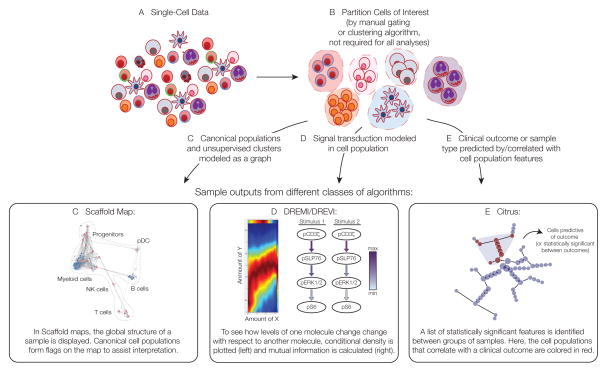
Technology development in biological research often aims to either increase the number of cellular features that can be surveyed simultaneously or enhance the resolution at which such observations are possible. For decades, flow cytometry has balanced these goals to fill a critical need by enabling the measurement of multiple features in single cells, commonly to examine complex or hierarchical cellular systems. Recently, a format for flow cytometry has been developed that leverages the precision of mass spectrometry. This fusion of the two technologies, termed mass cytometry, provides measurement of over 40 simultaneous cellular parameters at single-cell resolution, significantly augmenting the ability of cytometry to evaluate complex cellular systems and processes. In this Primer, we review the current state of mass cytometry, providing an overview of the instrumentation, its present capabilities, and methods of data analysis, as well as thoughts on future developments and applications.


Figures

Figure 1
Figure 1. Workflow of a typical mass cytometry experiment
Single cells are acquired, and a viability stain is applied to mark dead cells for exclusion from analyses. Fixation can optionally be applied at this point to preserve the cell state. Multiple samples can be barcoded with unique combinations of heavy metal tags, enabling them to be pooled together prior to staining to minimize technical variability at this step. After pooling samples into one tube, cells are then incubated with antibodies targeted against proteins of interest. Cell permeabilization can be performed if intracellular targets are to be measured. Cells are nebulized into droplets as they are introduced into the mass cytometer. They then travel into an inductively-coupled argon plasma (ICP), in which covalent bonds are broken and ions are liberated. The ion cloud is filtered by a quadrupole to remove common biological elements and enrich the heavy metal reporter ions to be quantified by time-of-flight mass spectrometry. Ion signals are integrated on a per-cell basis, resulting in single-cell measurements for downstream analysis. Data are compiled in an FCS file that can then be parsed and plotted in a variety of ways.
Figure 2
Figure 2. Computational methods developed for mass cytometry data analysis
Several classes of tools have been recently developed to assist in the interpretation of mass cytometry data. Here, we focus on those methods developed specifically for this purpose. Sample results are shown from different classes of algorithms designed to assess the global structure of a sample (Scaffold maps), the relationship between two molecules in single cells (DREMI/DREVI), or the cellular/molecular features that correlate with or best predict a clinical outcome or sample type (Citrus), respectively.
Figure 3
Figure 3. The challenge of visualizing high-dimensional data without information loss
High dimensional data is challenging to visualize due to the amount of information that must be captured in an effective representation. Here are two examples of how information content is lost by compressing the dimensionality of data. A) 2-dimensional data is compressed into 1 dimension: Left) the frequency of a property called dimension 2 is plotted against the frequency of a property called dimension 1, revealing 3 distinct cell populations. Right) Plotting just the frequency of the Dimension 1 property loses this view. B) 3-dimensional data showing the range of cells that have 3 different properties (dimensions) and how they relate to each other (left) is compressed into 2 dimensions (middle) and 1 dimension (right). The trajectory of cells is entirely lost in the lower-dimensional representations.

References

    1. Aghaeepour N, Finak G, Hoos H, Mosmann TR, Brinkman R, Gottardo R, Scheuermann RH FlowCAP Consortium, DREAM Consortium. Critical assessment of automated flow cytometry data analysis techniques. Nature Methods. 2013;10:228–238. - PMC - PubMed
    1. Amir E-AD, Davis KL, Tadmor MD, Simonds EF, Levine JH, Bendall SC, Shenfeld DK, Krishnaswamy S, Nolan GP, Pe’er D. viSNE enables visualization of high dimensional single-cell data and reveals phenotypic heterogeneity of leukemia. Nature Biotechnology 2013 - PMC - PubMed
    1. Angelo M, Bendall SC, Finck R, Hale MB, Hitzman C, Borowsky AD, Levenson RM, Lowe JB, Liu SD, Zhao S, et al. Multiplexed ion beam imaging of human breast tumors. Nat Med. 2014;20:436–442. - PMC - PubMed
    1. Bair E, Tibshirani R. Semi-supervised methods to predict patient survival from gene expression data. PLoS Biol. 2004;2:e108. - PMC - PubMed
    1. Bandura DR, Baranov VI, Ornatsky OI, Antonov A, Kinach R, Lou X, Pavlov S, Vorobiev S, Dick JE, Tanner SD. Mass cytometry: technique for real time single cell multitarget immunoassay based on inductively coupled plasma time-of-flight mass spectrometry. Anal Chem. 2009;81:6813–6822. - PubMed