. 2016 May 5;62(3):385-396.
doi: 10.1016/j.molcel.2016.04.003.

Inhibition of DHHC20-Mediated EGFR Palmitoylation Creates a Dependence on EGFR Signaling

Affiliations

Inhibition of DHHC20-Mediated EGFR Palmitoylation Creates a Dependence on EGFR Signaling

Kristin B Runkle et al. Mol Cell. .

Abstract

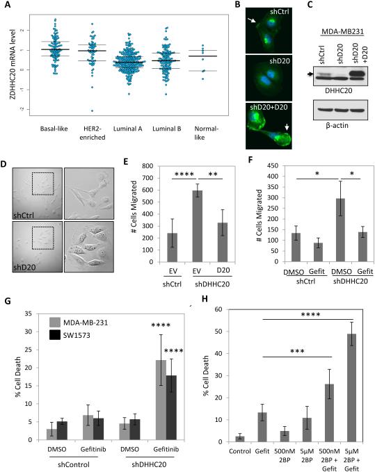
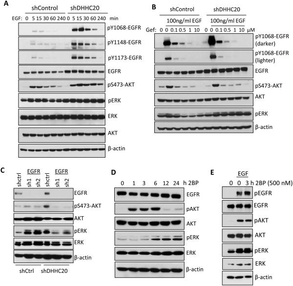
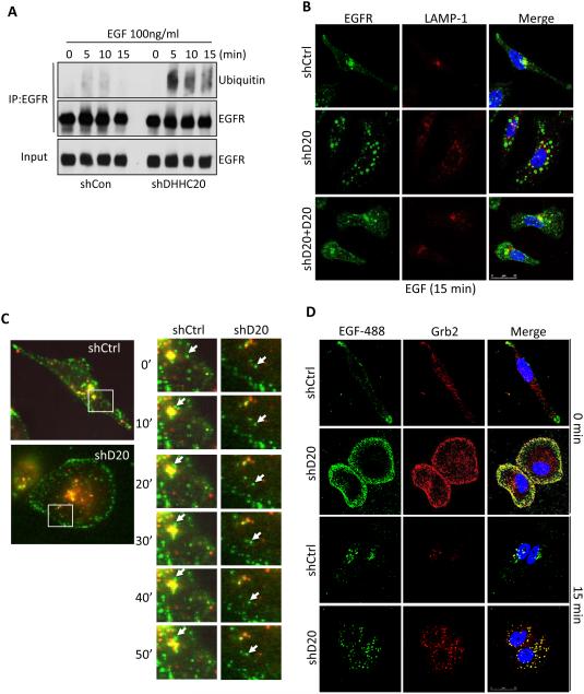
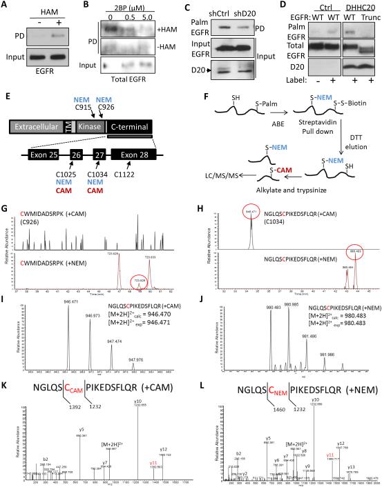
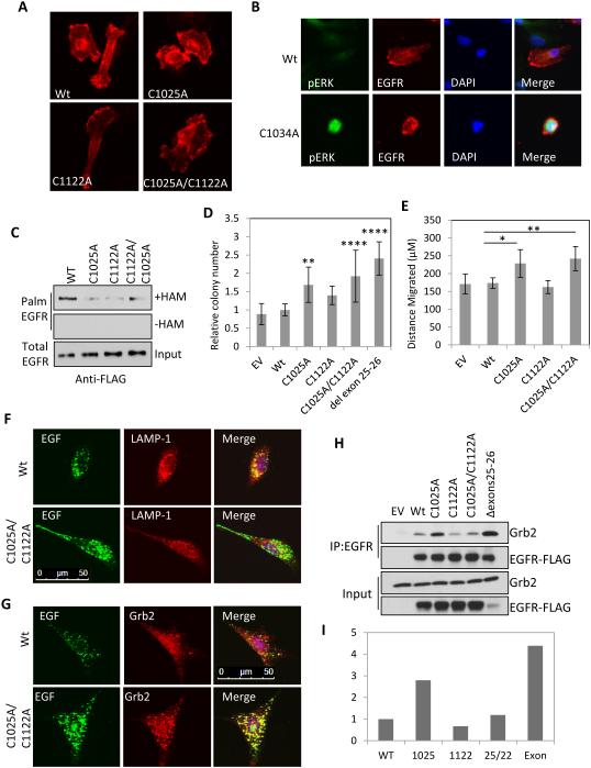
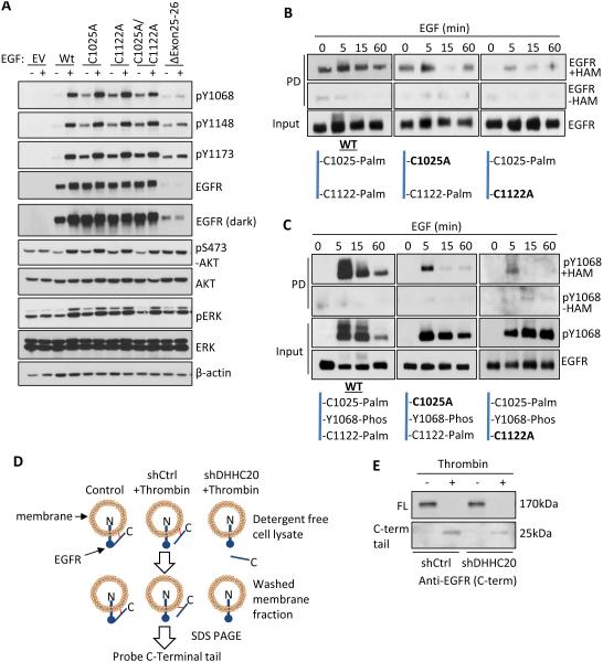
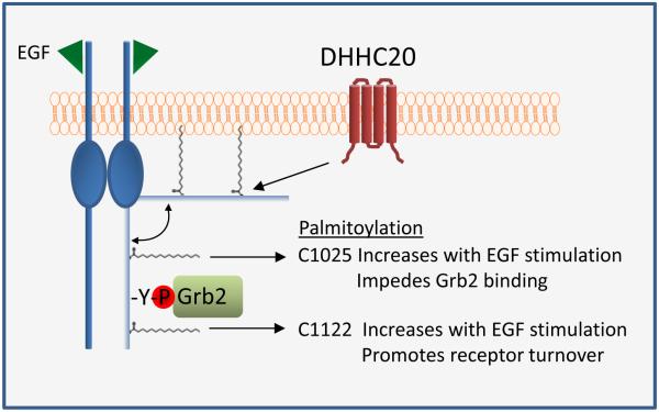
Inappropriate activation of the receptor tyrosine kinase EGFR contributes to a variety of human malignancies. Here we show a mechanism to induce vulnerability to an existing first line treatment for EGFR-driven cancers. We find that inhibiting the palmitoyltransferase DHHC20 creates a dependence on EGFR signaling for cancer cell survival. The loss of palmitoylation increases sustained EGFR signal activation and sensitizes cells to EGFR tyrosine kinase inhibition. Our work shows that the reversible modification of EGFR with palmitate "pins" the unstructured C-terminal tail to the plasma membrane, impeding EGFR activation. We identify by mass spectrometry palmitoylated cysteine residues within the C-terminal tail where mutation of the cysteine residues to alanine is sufficient to activate EGFR signaling promoting cell migration and transformation. Our results reveal that the targeting of a peripheral modulator of EGFR signaling, DHHC20, causes a loss of signal regulation and susceptibility to EGFR inhibitor-induced cell death.


Figures

Figure 1
Figure 1. Silencing DHHC20 increases EGFR-dependent cell migration and enhances Gefitinib-induced cytotoxicity
(A) DHHC20 mRNA expression is altered in human breast cancer subtypes. Global patterns of ZDHHC20 expression were identified in human breast invasive carcinoma in the TCGA database; Basal-like (n=98), HER2-enriched (n=58), Luminal A (n=230), Luminal B (n=125) and Normal-like (n=8). (B-C) DHHC20 expression measured by immunoblotting and immunofluorescence microscopy is inhibited by shRNA in MDA-MB-231 cells. Expression of shRNA resistant DHHC20 produces a product with the same molecular weight and cellular localization as the endogenous protein. (B) Immunofluorescence staining of DHHC20 (green) and DAPI (blue) show expression of DHHC20 at the plasma membrane (arrow) and perinuclear region. (C) Immunoblotting with a DHHC20 specific antibody shows inhibition of the DHHC20 band (arrow). (D) Silencing DHHC20 in MDA-MB-231 cells induces cell spreading with increased membrane ruffling. (E) Knockdown of DHHC20 increases chemotaxis towards FBS and this is rescued by expression of shRNA resistant DHHC20 (mean +/−StDev). (F) Increased chemotaxis of shDHHC20 MDA-MB-231 cells is inhibited by treatment with Gefitinib (10μM) (mean +/−StDev). (G) Gefitinib treatment (10 μM) increases cytotoxicity in DHHC20 silenced MDA-MB-231 (gray bars) and SW1573 (black bars) cells (mean +/−StDev). (H) Inhibiting palmitoylation with 2BP increases Gefitinib induced cytotoxicity. MDA-MB-231 cells were treated with 10μM Gefitinib alone or in combination with 500nM or 5μM 2BP (mean +/−StDev).
Figure 2
Figure 2. DHHC20 knockdown increases EGFR activation and signaling
(A) Silencing DHHC20 in MDA-MB-231 cells increases EGFR expression and EGF (100ng/ml) induced phosphoryation of EGFR and AKT. (B) Gefitinib inhibits activation of EGFR and AKT. MDA-MB-231 cells were serum starved in the presence of 10μM Gefitinib, treated with EGF (100ng/ml) for 15 minutes and activation of EGFR, AKT, and ERK was determined by SDS-PAGE. (C) The increased phosphorylation of ERK in DHHC20 silenced MDA-MB-231 cells is inhibited by EGFR shRNA. (D) MDA-MB-231 cells were treated with 500nM 2BP and the activation of AKT and ERK was analyzed by SDS-PAGE. (E) 2BP increases EGF-induced EGFR activation in MDA-MB-231 cells treated with 500nM 2BP for 3 hours and then stimulated with EGF for15 minutes.
Figure 3
Figure 3. Silencing DHHC20 expression disrupts EGFR endocytic trafficking
(A) Ubiquitination of EGFR is increased in shDHHC20 MDA-MB-231 cells treated with EGF (100ng/ml) and EGFR was immunoprecipitated using anti-EGFR (sc-120). (B) Silencing DHHC20 decreases EGF-induced EGFR (green) trafficking to LAMP-1 (red) positive lysosomes, which are rescued by exogenous expression of DHHC20. DAPI (blue). Colocalization of EGFR and LAMP-1 was determined by Mander’s Overlap Coefficient; shControl (0.403), shDHHC20 (0.242), shDHHC20+DHHC20 (0.395). (C) Live cell imaging shows that trafficking of Alexa-fluor488 labeled EGF (green) to lysosomes (red) is disrupted in shDHHC20 MDA-MB-231 cells. (D) Silencing DHHC20 increases Grb2 (red) localization to Alexa-fluor488 labeled EGF (green) labelled endosomes in MDA-MB-231 DAPI (blue). Colocalization between EGFR and LAMP-1 was determined by Mander’s Overlap Coefficient; shControl (0.627), shDHHC20 (0.723).
Figure 4
Figure 4. DHHC20 palmitoylates EGFR within the C-terminal tail
(A) EGFR palmitoylation in MDA-MB-231 cells determined using an acyl-biotinyl exchange (ABE) assay. (B) The palmitoyltransferase inhibitor 2BP reduces EGFR palmitoylation. MDA-MB-231 cells were treated with 2BP for 24 hours and EGFR palmitoylation was determined by ABE. (C) Silencing DHHC20 decreases EGFR palmitoylation in MDA-MB-231 cells determined by metabolic labeling. (D) DHHC20 palmitoylates the C-terminal tail of EGFR. HEK293T cells transiently expressing full length EGFR (WT) or a C-terminal tail truncation mutant (Trunc) and either empty vector control (EV) or DHHC20. (E) Schematic of EGFR including the exons and cysteine residues located within the C-terminal tail and kinase domain. Numbering corresponds to human EGFR excluding the signal sequence. (F) Experimental strategy for detecting EGFR palmitoylation by mass spectrometry. (G) Selected ion chromatograms of the [M+2H]2+ peptide (CWMIDADSRPK) with both potential carbamidomethylation (CAM, +57 Da) and N-Ethylmaleimide (NEM, +125 Da) modifications of cysteine 926. As can be seen in the chromatograms, only a peak for the CWMIDADSRPK (+NEM) peptide was found, and no peaks for CWMIDADSRPK (+CAM) were observed. (H) Selected ion chromatograms of the [M+2H]2+ peptide (NGLQSCPIKEDSFLQR) with both potential carbamidomethylation (CAM, +57 Da) and N-Ethylmaleimide (NEM, +125 Da) modifications of cysteine 1034. As can be seen in the chromatograms, both peaks for NGLQSCPIKEDSFLQR (+NEM) and NGLQSCPIKEDSFLQR (+CAM) were observed (~ 10 min retention time shift between the species). (I) Full mass spectrum of the parent ion corresponding to the NGLQSCPIKEDSFLQR (+CAM), and accurate mass confirms the correct assignment. (J) Full mass spectrum of the parent ion corresponding to the NGLQSCPIKEDSFLQR (+NEM), and accurate mass confirms the correct assignment. (K) MS/MS spectrum of the [M+2H]2+ peptide NGLQSCPIKEDSFLQR (+CAM), with highlighted fragment ion indicating the CAM modification on the Cys residue 1034. (L) MS/MS spectrum of the [M+2H]2+ peptide NGLQSCPIKEDSFLQR (+NEM), with highlighted fragment ion indicating the NEM modification on the cysteine residue 1034.
Figure 5
Figure 5. Mutation in C1025 and C1122 activates EGFR, promotes cellular transformation and increases cell migration
(A) Mutation of cysteine residues 1025 and 1122 of EGFR-FLAG (red) does not alter receptor localization in MDA-MB-231 cells. (B) Expression of C1034A mutant EGFR-FLAG (red) in MDA-MB-231 cells increases ERK phosphorylation (green), DAPI (blue). (C) Mutation of EGFR cysteine residues 1025, 1122 and 1025/1122 decreases EGFR palmitoylation in HEK293T cells. (D) Mutation of EGFR C1025, C1025/C1122 or deletion of exons 25-26 deletion increases colony formation of NIH 3T3 cells grown in soft agar. Expression levels of each mutant are shown in Figure S7A (mean +/− StDev). (E) Mutation of EGFR C1025 and C1025/C1122 increases migration of NIH 3T3 cells (mean +/− SEM). (F) Mutation of EGFR C1025/C1122 delays EGFR endocytosis and trafficking of EGF-alexafluor488 (green) labelled endosomes to lysosomes stained for LAMP-1 (red). Cells expressing the EGFR C1025/C1122 mutant have increased localization of Grb2 (red) to EGF-alexafluor488 (green) labelled endosomes. (H) The C1025A mutation or the exon 25-26 deletion increases the interaction of EGFR-FLAG with endogenous Grb2 in HEK293T cells. EGFR was immunoprecipitated with anti-EGFR (sc-120 AF488). (I) Quantification of co-immunopreciptated Grb2 by densitometric analysis of immunoblots in (H). Y-axis indicates arbitrary units normalized to WT EGFR.
Figure 6
Figure 6. Palmitoylation regulates the activity of EGFR in response to EGF
(A) Mutation of EGFR palmitoylated cysteine residues increases EGFR activation independent of EGF in NIH 3T3 cells compared to WT EGFR. (B) EGFR palmitoylation increases upon EGF (100ng/ml) stimulation in MDA-MB-231 cells. (C) Dually phosphorylated and palmitoylated EGFR and EGFR mutants C1025A and C1122A turns over more rapidly than total EGFR in response to EGF (100ng/ml). (D) Experimental strategy of the thrombin cleavage assay. (E) Palmitoylation pins the C-terminal tail of EGFR to the cell membrane. HEK293T cells transiently expressing EGFR-FLAG containing a thrombin cleavage site (LVPRGS) at Gly959 were lysed in the presence or absence of thrombin protease. The membrane fraction was isolated by centrifugation and EGFR was immunoprecipitated from the membrane fraction. The presence of membrane bound full length (170kDa) EGFR and the cleaved C-terminal tail (25kDa) was determined by SDS-PAGE.
Figure 7
Figure 7. Mechanistic summary of the effects of palmitoylation on EGFR activation and signaling
Palmitoylation of the C-terminus of EGFR promotes membrane association. Palmitoylation of EGFR at C1025 impedes the binding of Grb2 to EGFR whereas palmitoylation at C1122 increases with EGF stimulation and promotes receptor turnover.

References

    1. Aicart-Ramos C, Valero RA, Rodriguez-Crespo I. Protein palmitoylation and subcellular trafficking. Biochimica et biophysica acta. 2011;1808:2981–2994. - PubMed
    1. Bollu LR, Katreddy RR, Blessing AM, Pham N, Zheng B, Wu X, Weihua Z. Intracellular activation of EGFR by fatty acid synthase dependent palmitoylation. Oncotarget. 2015;6:34992–35003. - PMC - PubMed
    1. Cho J, Pastorino S, Zeng Q, Xu X, Johnson W, Vandenberg S, Verhaak R, Cherniack AD, Watanabe H, Dutt A, et al. Glioblastoma-derived epidermal growth factor receptor carboxyl-terminal deletion mutants are transforming and are sensitive to EGFR-directed therapies. Cancer research. 2011;71:7587–7596. - PMC - PubMed
    1. Conibear E, Davis NG. Palmitoylation and depalmitoylation dynamics at a glance. Journal of cell science. 2010;123:4007–4010. - PMC - PubMed
    1. Ekstrand AJ, Sugawa N, James CD, Collins VP. Amplified and rearranged epidermal growth factor receptor genes in human glioblastomas reveal deletions of sequences encoding portions of the N- and/or C-terminal tails. Proceedings of the National Academy of Sciences of the United States of America. 1992;89:4309–4313. - PMC - PubMed

MeSH terms