. 2016 May 6:15:75.
doi: 10.1186/s12933-016-0383-8.

Oral treatment with a zinc complex of acetylsalicylic acid prevents diabetic cardiomyopathy in a rat model of type-2 diabetes: activation of the Akt pathway

Affiliations

Oral treatment with a zinc complex of acetylsalicylic acid prevents diabetic cardiomyopathy in a rat model of type-2 diabetes: activation of the Akt pathway

Sevil Korkmaz-Icöz et al. Cardiovasc Diabetol. .

Abstract

Background: Type-2 diabetics have an increased risk of cardiomyopathy, and heart failure is a major cause of death among these patients. Growing evidence indicates that proinflammatory cytokines may induce the development of insulin resistance, and that anti-inflammatory medications may reverse this process. We investigated the effects of the oral administration of zinc and acetylsalicylic acid, in the form of bis(aspirinato)zinc(II)-complex Zn(ASA)2, on different aspects of cardiac damage in Zucker diabetic fatty (ZDF) rats, an experimental model of type-2 diabetic cardiomyopathy.

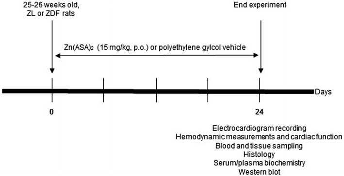
Methods: Nondiabetic control (ZL) and ZDF rats were treated orally with vehicle or Zn(ASA)2 for 24 days. At the age of 29-30 weeks, the electrical activities, left-ventricular functional parameters and left-ventricular wall thicknesses were assessed. Nitrotyrosine immunohistochemistry, TUNEL-assay, and hematoxylin-eosin staining were performed. The protein expression of the insulin-receptor and PI3K/AKT pathway were quantified by Western blot.

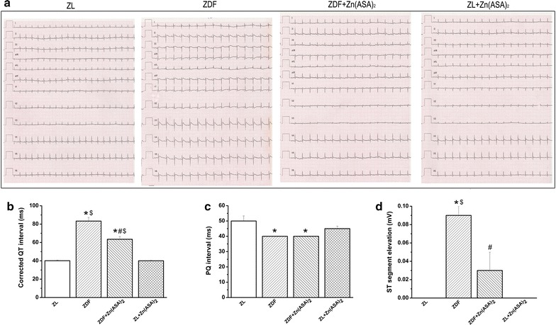
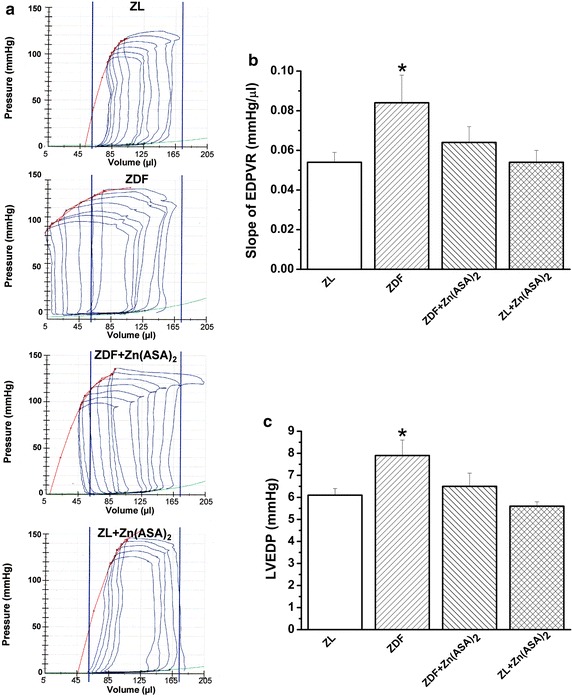
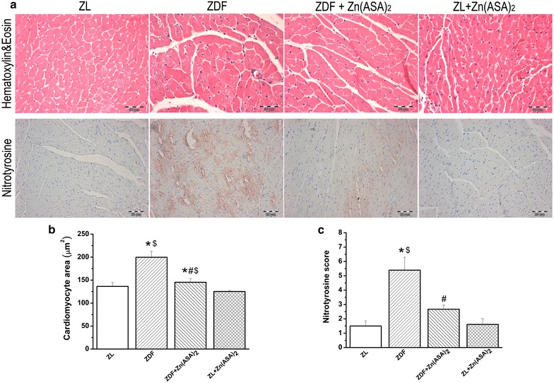
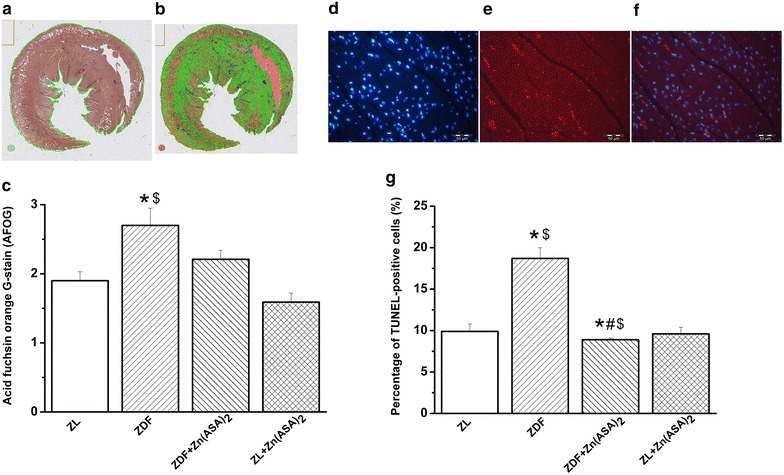
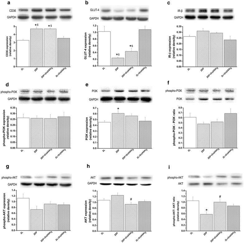
Results: Zn(ASA)2-treatment significantly decreased plasma glucose concentration in ZDF rats (39.0 ± 3.6 vs 49.4 ± 2.8 mM, P < 0.05) while serum insulin-levels were similar among the groups. Data from cardiac catheterization showed that Zn(ASA)2 normalized the increased left-ventricular diastolic stiffness (end-diastolic pressure-volume relationship: 0.064 ± 0.008 vs 0.084 ± 0.014 mmHg/µl; end-diastolic pressure: 6.5 ± 0.6 vs 7.9 ± 0.7 mmHg, P < 0.05). Furthermore, ECG-recordings revealed a restoration of prolonged QT-intervals (63 ± 3 vs 83 ± 4 ms, P < 0.05) with Zn(ASA)2. Left-ventricular wall thickness, assessed by echocardiography, did not differ among the groups. However histological examination revealed an increase in the cardiomyocytes' transverse cross-section area in ZDF compared to the ZL rats, which was significantly decreased after Zn(ASA)2-treatment. Additionally, a significant fibrotic remodeling was observed in the diabetic rats compared to ZL rats, and Zn(ASA)2-administered ZDF rats showed a similar collagen content as ZL animals. In diabetic hearts Zn(ASA)2 significantly decreased DNA-fragmentation, and nitro-oxidative stress, and up-regulated myocardial phosphorylated-AKT/AKT protein expression. Zn(ASA)2 reduced cardiomyocyte death in a cellular model of oxidative stress. Zn(ASA)2 had no effects on altered myocardial CD36, GLUT-4, and PI3K protein expression.

Conclusions: We demonstrated that treatment of type-2 diabetic rats with Zn(ASA)2 reduced plasma glucose-levels and prevented diabetic cardiomyopathy. The increased myocardial AKT activation could, in part, help to explain the cardioprotective effects of Zn(ASA)2. The oral administration of Zn(ASA)2 may have therapeutic potential, aiming to prevent/treat cardiac complications in type-2 diabetic patients.

Keywords: Cardiac function; Diabetic cardiomyopathy; Type-2 diabetes mellitus; Zinc-aspirin complex.


Figures

Fig. 1
Fig. 1
Experimental protocol. At the age of 25–26 week-old, ZL or ZDF rats were treated with Zn(ASA)2 or a polyethylene glycol vehicle (15 mg/kg) orally for 24 consecutive days. Zn(ASA) 2 indicates a zinc complex of acetylsalicylic acid, ZDF Zucker diabetic fatty rats, ZL Zucker lean
Fig. 2
Fig. 2
Total cholesterol and triglyceride levels of fast performance liquid chromatography (FPLC) fractions. The graph shows a cholesterol and b triglyceride concentrations of FPLC fractions from pooled serum. Zn(ASA) 2 indicates a zinc complex of acetylsalicylic acid, ZDF Zucker diabetic fatty rats, ZL Zucker lean, VLDL very low density lipoprotein, LDL low density lipoprotein, HDL high density lipoprotein
Fig. 3
Fig. 3
Zn(ASA)2 improves electrocardiographic pattern. a Representative surface 12-lead ECG tracing; b corrected QT interval; c PQ-interval, and d ST-segment elevation. Values are mean ± SEM, n = 6-10. *P < 0.05 vs ZL, #P < 0.05 vs ZDF, $P < 0.05 vs Zn(ASA)2, nQTc = QT/(RR/f)1/2. Zn(ASA) 2 indicates a zinc complex of acetylsalicylic acid, ZDF Zucker diabetic fatty rats, ZL Zucker lean, RR corresponding to cardiac cycle length and f to frequency, nQTc QT intervals corrected with RR intervals
Fig. 4
Fig. 4
Zn(ASA)2 improves left-ventricular cardiac function. a Representative original pressure–volume loops registered during transient occlusion of the inferior vena cava. The slope of end-systolic pressure–volume relationship (red line) and end-diastolic pressure–volume relationship (EDPVR, green line); b the slope of end-diastolic pressure–volume relationship (EDPVR); and c left-ventricular end-diastolic pressure (LVEDP). Values are mean ± SEM, n = 6–10. *P < 0.05 vs ZL, #P < 0.05 vs ZDF, $P < 0.05 vs Zn(ASA)2. Zn(ASA) 2 indicates a zinc complex of acetylsalicylic acid, ZDF Zucker diabetic fatty rats, ZL Zucker lean
Fig. 5
Fig. 5
Zn(ASA)2 decreases cardiomyocyte hypertrophy and nitro-oxidative stress. a Hematoxylin and eosin staining micrographs of transverse sections of myocardium (magnification ×400; scale bar: 50 μm) and representative photomicrographs of nitrotyrosine immunohistochemistry staining; magnification ×200, scale bar: 20 µm. b Quantitative analysis of cardiomyocyte cross-sectional area using of ~20 cardiomyocytes in each group and c immunohistochemical scores for nitrotyrosine in the myocardium. Values are mean ± SEM, n = 6–9. *P < 0.05 vs ZL, #P < 0.05 vs ZDF, $P < 0.05 vs Zn(ASA)2. Zn(ASA) 2 indicates a zinc complex of acetylsalicylic acid, ZDF Zucker diabetic fatty rats, ZL Zucker lean
Fig. 6
Fig. 6
Effect of Zn(ASA)2 on myocardial fibrosis and DNA-strand breaks in cardiomyocytes. Representative photomicrographs showing the whole-slide after a the region of interest detection and b pixelwise classification of the tissue; blue fibrotic areas, green normal tissue, orange excluded areas in the acid fuchsin orange G (AFOG) stained sections. c Quantitative analysis of interstitial fibrosis in the myocardium. Representative photomicrographs of d nuclei with 4′,6-diamidino-2phenylindole (DAPI-stained nuclei, blue); e nuclei with fragmented DNA visualized by Terminal Deoxynucleotidyl Transferase-Mediated dUTP Nick End-Labeling (TUNEL) staining (TUNEL-positive nuclei, red), and f merged image (red/blue double stained) (magnification ×400, scale bar: 50 µm) from the same sample which belong to the ZDF group. g Quantification of TUNEL-positive cells for each group. Values are mean ± SEM, n = 6–10. *P < 0.05 vs ZL, #P < 0.05 vs ZDF, $P < 0.05 vs Zn(ASA)2. Zn(ASA) 2 indicates a zinc complex of acetylsalicylic acid, ZDF Zucker diabetic fatty rats, ZL Zucker lean
Fig. 7
Fig. 7
Effects of Zn(ASA)2 on myocardial protein expression. Immunoblot analysis for a CD36, b insulin receptor (IR)-β, c glucose transporter (GLUT)-4, d phosphorylated PI3K, e PI3K, f phosphorylated phosphatidylinositol 3-kinase-(PI3K)/PI3K ratio, g phosphorylated, h total AKT and i phosphorylated AKT/total Akt ratio protein band densities in the myocardium. Glyceraldehyde-3-phosphate dehydrogenase (GAPDH), housekeeping protein was used as reference. Values are mean ± SEM, n = 6–7. *P < 0.05 vs ZL, #P < 0.05 vs ZDF, $P < 0.05 vs Zn(ASA)2. Zn(ASA) 2 indicates a zinc complex of acetylsalicylic acid, ZDF Zucker diabetic fatty rats, ZL Zucker lean

References

    1. Aneja A, Tang WH, Bansilal S, Garcia MJ, Farkouh ME. Diabetic cardiomyopathy: insights into pathogenesis, diagnostic challenges, and therapeutic options. Am J Med. 2008;121:748–757. doi: 10.1016/j.amjmed.2008.03.046. - DOI - PubMed
    1. Regan TJ, Lyons MM, Ahmed SS, Levinson GE, Oldewurtel HA, Ahmad MR, et al. Evidence for cardiomyopathy in familial diabetes mellitus. J Clin Invest. 1977;60:884–899. doi: 10.1172/JCI108843. - DOI - PMC - PubMed
    1. Fein FS. Diabetic cardiomyopathy. Diabetes Care. 1990;13:1169–1179. doi: 10.2337/diacare.13.11.1169. - DOI - PubMed
    1. Schaffer JE. Lipotoxicity: when tissues overeat. Curr Opin Lipidol. 2003;14:281–287. doi: 10.1097/00041433-200306000-00008. - DOI - PubMed
    1. Unger RH, Orci L. Diseases of liporegulation: new perspective on obesity and related disorders. Faseb J. 2001;15:312–321. doi: 10.1096/fj.00-0590. - DOI - PubMed

Publication types

MeSH terms