Review
. 2016 Jul;73(14):2707-25.
doi: 10.1007/s00018-016-2246-6. Epub 2016 May 6.

Remarkable evolutionary relatedness among the enzymes and proteins from the α-amylase family

Affiliations
Review

Remarkable evolutionary relatedness among the enzymes and proteins from the α-amylase family

Štefan Janeček et al. Cell Mol Life Sci. 2016 Jul.

Abstract

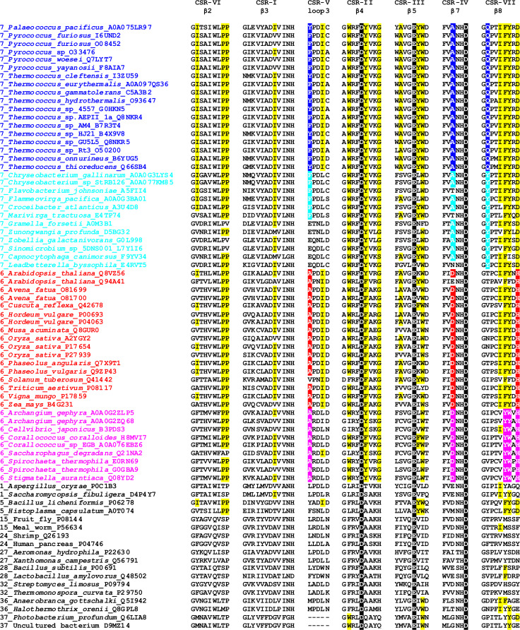
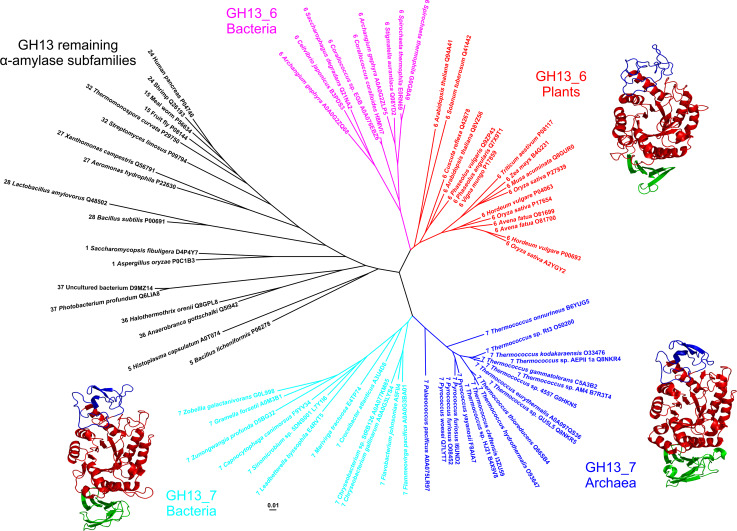
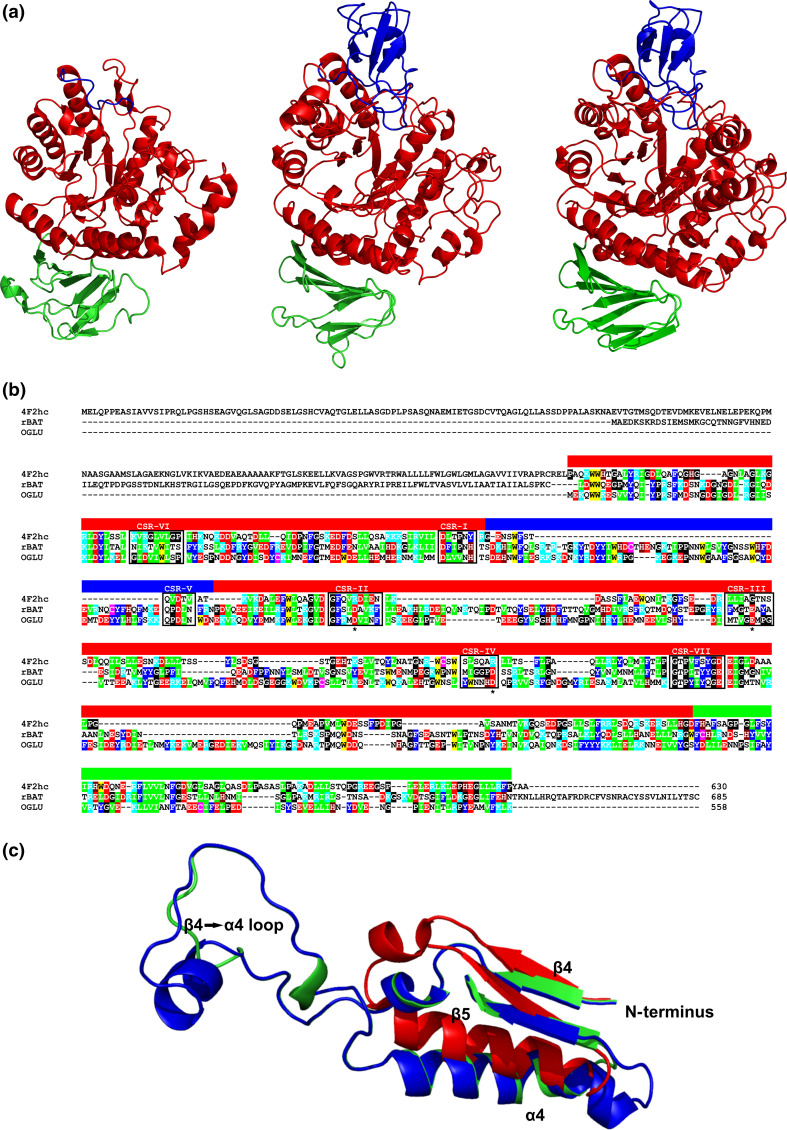
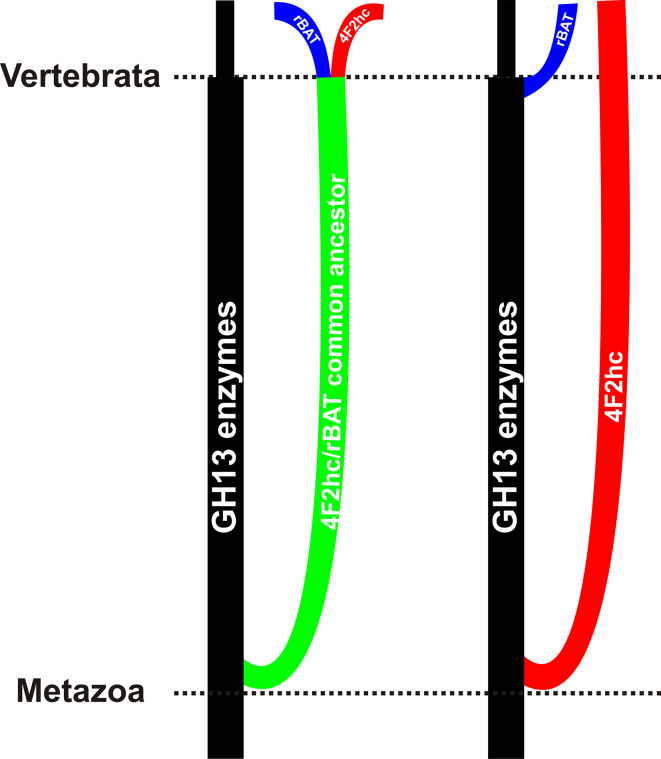
The α-amylase is a ubiquitous starch hydrolase catalyzing the cleavage of the α-1,4-glucosidic bonds in an endo-fashion. Various α-amylases originating from different taxonomic sources may differ from each other significantly in their exact substrate preference and product profile. Moreover, it also seems to be clear that at least two different amino acid sequences utilizing two different catalytic machineries have evolved to execute the same α-amylolytic specificity. The two have been classified in the Cabohydrate-Active enZyme database, the CAZy, in the glycoside hydrolase (GH) families GH13 and GH57. While the former and the larger α-amylase family GH13 evidently forms the clan GH-H with the families GH70 and GH77, the latter and the smaller α-amylase family GH57 has only been predicted to maybe define a future clan with the family GH119. Sequences and several tens of enzyme specificities found throughout all three kingdoms in many taxa provide an interesting material for evolutionarily oriented studies that have demonstrated remarkable observations. This review emphasizes just the three of them: (1) a close relatedness between the plant and archaeal α-amylases from the family GH13; (2) a common ancestry in the family GH13 of animal heavy chains of heteromeric amino acid transporter rBAT and 4F2 with the microbial α-glucosidases; and (3) the unique sequence features in the primary structures of amylomaltases from the genus Borrelia from the family GH77. Although the three examples cannot represent an exhaustive list of exceptional topics worth to be interested in, they may demonstrate the importance these enzymes possess in the overall scientific context.

Keywords: Evolutionary relatedness; Family GH77 amylomaltases of borrelian origin; Heavy-chains of rBAT and 4F2 proteins; Plant and archaeal α-amylases; α-Amylase family GH13.


Figures

Fig. 1
Fig. 1
Comparison of seven CSRs of the family GH13 α-amylases with focus on the subfamilies GH13_6 and GH13_7. These CSRs cover mostly individual β-strands of the catalytic TIM-barrel domain [32]. Each α-amylase in the list is characterized by the GH13 subfamily number [46], the source of origin (the organism) and the accession number from the UniProt database [221]. The α-amylases from the subfamilies GH13_6 and GH13_7 were collected from the actual CAZy database [37], whereas the set of representative α-amylases from the GH13 remaining subfamilies was prepared according to previous in silico studies [–79]. The catalytic triad is signified by black-and-white inversion. Sequence features characteristic of the GH13_7 archaeal α-amylases are highlighted in yellow. The features typical specifically for each group of α-amylases within the two GH13 subfamilies, i.e., (1) archaea and (flavo)-bacteria within GH13_7; and (2) plants and bacteria within GH13_6, which may discriminate the individual groups from each other are emphasized by respective colors
Fig. 2
Fig. 2
Evolutionary tree of the family GH13 α-amylases with focus on the subfamilies GH13_6 and GH13_7. The tree is based on the alignment of seven conserved sequence regions, shown in Fig. 1. The tree was calculated using the neighbor-joining clustering [222] implemented in the Clustal-W2 phylogeny package [223] available at the European Bioinformatics Institute’s web-site (http://www.ebi.ac.uk/), and then displayed with the program iTOL [224]. Tertiary structures of representatives of flavobacterial, archeal and plant family GH13 α-amylases are shown near their clusters in the tree. Sources of the α-amylases: Sinomicrobium sp. 5DNS001 [81] (subfamily GH13_7; flavobacteria); Pyrococcus woesei [72] (subfamily GH13_7; archaea); Hordeum vulgare—barley high pI isozyme AMY-2 [61] (subfamily GH13_6; plants). The archaeal and plant α-amylases are experimentally solved crystal structures retrieved from the Protein Data Bank (PDB) [225] under the PDB codes 1MWO and 1AMY, respectively. The flavobacterial α-amylase is a tertiary structure model obtained at the fold recognition server Phyre-2 [89] for its amino acid sequence (UniProt accession number: L7Y1I6; residues: Gly52-Gly477) based on the P. woesei α-amylase structure (1MWO) as template. The individual domains are colored as follows: catalytic (β/α)8-barrel—red, domain B—blue, domain C—green. The structural models were displayed with the program PyMol [226]
Fig. 3
Fig. 3
a Comparison of tertiary structures of the family GH13 hcHAT proteins from animals and α-glucosidase from bacteria. Sources of the proteins: Homo sapiens 4F2hc antigen [103] (subfamily GH13_34; left); Homo sapiens rBAT protein [111] (subfamily GH13_35; middle); Bacillus cereus oligo-1,6-glucosidase [96] (subfamily GH13_31; right). b Amino acid sequence alignment of the same three proteins: human 4F2hc antigen [103] (UniProt accession No.: P08195-1); human rBAT protein [111] (Q07837); Bacillus cereus oligo-1,6-glucosidase (OGLU) [96] (P21332). Color code for the selected residues: W, yellow; F, Y—blue; V, L, I—green; D, E—red; R, K—cyan; H—brown; C—magenta; G, P—black. The seven characteristic CSRs are boxed and marked above the alignment. The colored lane above the alignment blocks means the three domains shown in a. The catalytic triad is signified by asterisks under the alignment. c Structural overlay emphasizing the longer loop 4 connecting the strand β4 to the helix α4 in the oligo-1,6-glucosidase (green) and rBAT protein (blue) in comparison to a very short version present in the 4F2hc antigen (red). The structures were superimposed using the MultiProt web-server [227] (http://bioinfo3d.cs.tau.ac.il/MultiProt/); the overlap being characterized by 280 corresponding Cα-atoms and the RMSD value of 0.96 Å. The human 4F2hc antigen and bacterial oligo-1,6-glucosidase are experimentally solved crystal structures retrieved from the PDB [225] under the PDB codes 2DH3 and 1UOK, respectively. The human rBAT protein is a tertiary structure model obtained at the fold recognition server Phyre-2 [89] for its amino acid sequence (UniProt accession number: Q07837; residues: Asp116-Glu649) based on the B. cereus oligo-1,6-glucosidase structure (1UOK) as template. The individual domains are colored as follows: catalytic (β/α)8-barrel—red, domain B—blue, domain C—green. The structures were visualized with the program PyMol [226]
Fig. 4
Fig. 4
Evolutionary scenarios of hcHAT proteins with respect to the α-amylase family GH13. The left eventuality considers a single-event division of both rBAT proteins and 4F2hc antigens from the enzymes of the α-amylase family in basal Metazoa with a subsequent split to rBAT proteins and 4F2 antigens in higher animals like chordates. The right possibility counts with two independent evolutionary events leading first to separation of the 4F2hc antigens from the α-amylase family enzymes in the basal Metazoa and second to recruitment of the rBAT proteins from enzymatic members of the oligo-1,6-glucosidase subfamily in higher animals
Fig. 5
Fig. 5
Tertiary structures of the family GH77 amylomaltases from bacteria and their comparison. Sources of the amylomaltases: a Thermus aquaticus [139]; b Borrelia burgdorferi [129]. c Structural overlay focusing on the active-site residues with complexed acarbose in the Thermus amylomaltase (red) compared with the naturally mutated corresponding residues in the Borrelia amylomaltase (blue). The structures were superimposed using the MultiProt web-server [227] (http://bioinfo3d.cs.tau.ac.il/MultiProt/); the overlap being characterized by 488 corresponding Cα-atoms and the RMSD value of 0.18 Å. The Thermus amylomaltase is experimentally solved crystal structure retrieved from the PDB [225] under the PDB code 1ESW, whereas the Borrelia amylomaltase is a tertiary structure model obtained at the homology modeling server SwissModel [228] for its amino acid sequence (UniProt accession number: A6YM39; residues: Asn12-Ala507) based on the T. aquaticus amylomaltase structure (1CWY [44]) as template. The individual domains are colored as follows: catalytic (β/α)8-barrel—red, subdomain B1—cyan, subdomain B2—magenta, subdomain B3—green. The exact positions of displayed active-site residues in the amino acid sequence can be identified in the alignment shown in Fig. 6. Based on the accepted nomenclature [229] the acarbose occupies the subsites from −3 to +1. The structures were visualized with the program PyMol [226]
Fig. 6
Fig. 6
Amino acid sequence comparison of family GH77 amylomaltases from borreliae and Thermus aquaticus. The four amylomaltases from borrelian origin represent different subgroups within the genus Borrelia [122] exhibiting unique mutations in several important active-site positions with respect to a typical bacterial amylomaltase represented by the one from Thermus [129, 145]. The sequences were retrieved from the UniProt database [221] according to their accession numbers succeeding the name of the organism. The alignment was done using the program Clustal-Omega [230] available at the European Bioinformatics Institute’s web-site (http://www.ebi.ac.uk/). The unique borreliae-like positions (Asn228, Lys306 and Gly407) and the catalytic triad (Asp308, Glu355 and Asp408; B. burgdorferi amylomaltase numbering) are signified by yellow/red and blue highlighting, respectively. The positions in amylomaltases from borreliae identical to that from Thermus are shown as dots. The black highlighting in the T. aquaticus amylomaltase means that all four borrelian counterparts have the same residue in those positions. The positions that are signified by black highlighting in amylomaltases from borreliae are identical among them but different from those in the enzyme from Thermus. The seven CSRs known for the entire α-amylase clan GH-H are boxed and marked as CSR-I to CSR-VII with indicated well-accepted secondary structure elements [32]. The individual domains are indicated as a colored lane above the alignment blocks as follows: catalytic (β/α)8-barrel—red, subdomain B1—cyan, subdomain B2—magenta, subdomain B3—green

References

    1. Janecek S, Svensson B, MacGregor EA. α-Amylase—an enzyme specificity found in various families of glycoside hydrolases. Cell Mol Life Sci. 2014;71:1149–1170. doi: 10.1007/s00018-013-1388-z. - DOI - PMC - PubMed
    1. MacGregor EA. α-Amylase structure and activity. J Protein Chem. 1988;7:399–415. doi: 10.1007/BF01024888. - DOI - PubMed
    1. Vihinen M, Mäntsälä P. Microbial amylolytic enzymes. Crit Rev Biochem Mol Biol. 1989;24:329–418. doi: 10.3109/10409238909082556. - DOI - PubMed
    1. Pandey A, Nigam P, Soccol CR, Soccol VT, Singh D, Mohan R. Advances in microbial amylases. Biotechnol Appl Biochem. 2000;31:135–152. doi: 10.1042/BA19990073. - DOI - PubMed
    1. Søgaard M, Ji Abe, Martin-Eauclaire MF, Svensson B. α-Amylases: structure and function. Carbohydr Polym. 1993;21:137–146. doi: 10.1016/0144-8617(93)90008-R. - DOI

Publication types

LinkOut - more resources