Review
. 2016 Oct 1;105(Pt B):109-144.
doi: 10.1016/j.addr.2016.04.028. Epub 2016 May 3.

Toxicology of graphene-based nanomaterials

Affiliations
Review

Toxicology of graphene-based nanomaterials

Gaurav Lalwani et al. Adv Drug Deliv Rev. .

Abstract

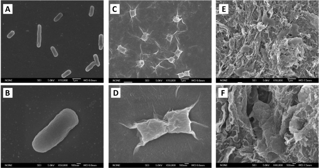
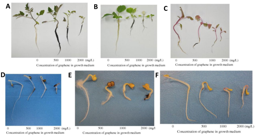
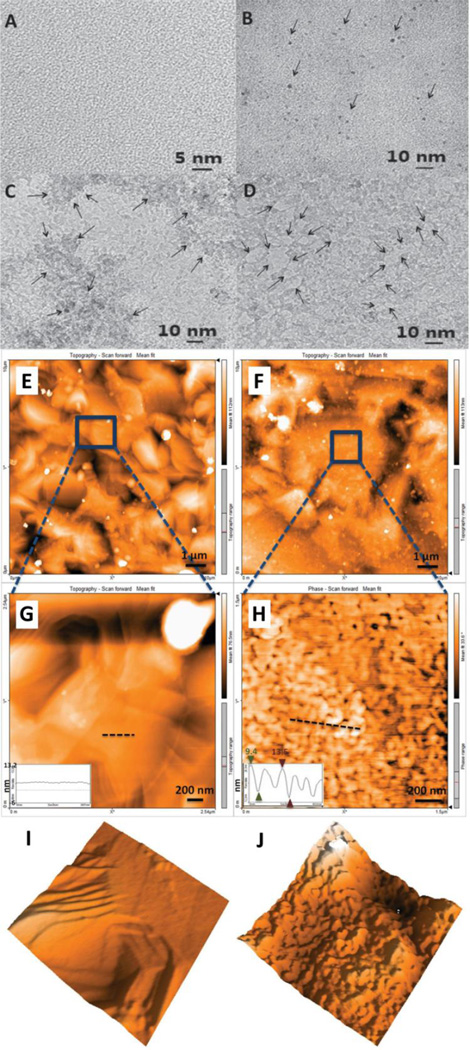
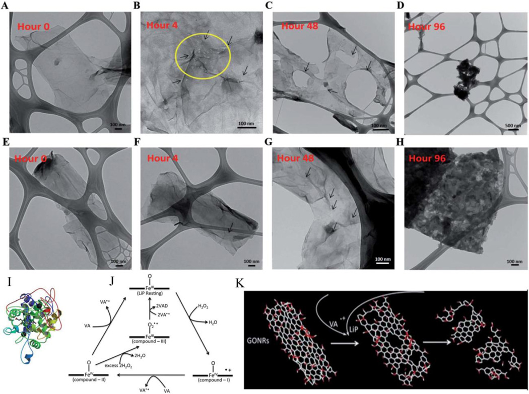
Graphene based nanomaterials possess remarkable physiochemical properties suitable for diverse applications in electronics, telecommunications, energy and healthcare. The human and environmental exposure to graphene-based nanomaterials is increasing due to advancements in the synthesis, characterization and large-scale production of graphene and the subsequent development of graphene based biomedical and consumer products. A large number of in vitro and in vivo toxicological studies have evaluated the interactions of graphene-based nanomaterials with various living systems such as microbes, mammalian cells, and animal models. A significant number of studies have examined the short- and long-term in vivo toxicity and biodistribution of graphene synthesized by variety of methods and starting materials. A key focus of these examinations is to properly associate the biological responses with chemical and morphological properties of graphene. Several studies also report the environmental and genotoxicity response of pristine and functionalized graphene. This review summarizes these in vitro and in vivo studies and critically examines the methodologies used to perform these evaluations. Our overarching goal is to provide a comprehensive overview of the complex interplay of biological responses of graphene as a function of their physiochemical properties.

Keywords: Antimicrobial; Biodistribution; Environmental; Graphene; In vitro; In vivo; Toxicity.


Figures

Figure 1
Figure 1
Graphene is the building material for 0D fullerenes, 1D carbon nanotubes and 3D graphite. Schematic adapted from Reference [6] with permission, copyright © Macmillan Publishers Limited, 2007.
Figure 2
Figure 2
Number of publications with the keyword ‘graphene’ from 1960–2015. Data retrieved from PubMed (www.ncbi.nlm.nih.gov).
Figure 3
Figure 3
Representative transmission electron microscopy images of (A and B) graphene nanoribbons, (C) graphene nanoplatelets, (D) graphene nanoonions, (E) graphene nanosheets and (F) graphene quantum dots. Image (A) adapted from Reference [41], (B–D) adapted from Reference [44], (E) adapted from Reference [96] and (F) adapted from Reference [146], with permissions. (A) copyright © American Chemical Society 2013, (B–D) copyright © Elsevier 2014, (E) copyright © Elsevier 2015, and (F) copyright © American Chemical Society, 2013.
Figure 4
Figure 4
Effects on (A) lactate dehydrogenase release, (B) reactive oxygen species generation and (C) caspase-3 activity (apoptosis marker) of PC12 cells treated with 0.1–100 µg/ml of graphene and single-walled carbon nanotubes. Adapted from Reference [61] with permission, copyright © American Chemical Society, 2010.
Figure 5
Figure 5
Representative transmission electron microscopy images of mesenchymal stem cells (MSC) treated with graphene nanoonions (GNOs, A&B) and oxidized-graphene nanoplatelets (GONPs, C&D) at 50 µg/ml for 24 hours. Yellow arrows correspond to aggregates of GNO visualized in vacuoles (green arrows). No nuclear uptake of GNOs was observed. Blue arrows correspond to aggregates of GONPs. GONPs were observed inside the nucleus (red arrows). Oil red O staining after adipogenic differentiation of MSC treated with 50 µg/ml of (E) GNO, (F) GONR and (G) GONP. Alizarin Red staining after osteogenic differentiation of MSC treated with 50 µg/ml of (H) GNO, (I) GONR and (J) GONP. No changes in the adipogenic and osteogenic differentiation of MSCs were observed. Adapted from Reference [44] with permission, copyright © Elsevier, 2014.
Figure 6
Figure 6
(A) Representative atomic force microscopy (AFM) image of graphene quantum dots (GQDs). Inset in image A depicts AFM height profile. (B) Cell viability of A549 cells assessed by WST-1 assay. Data reported as means ± SE. No significant differences in cell viability were observed upto a treatment concentration of 320 µg/ml. (C) Cell viability assessed by WST-1 assay, (D) cell apoptosis and necrosis (E) LDH assay and (F) ROS generation by HeLa cells upon treatment with 0–160 µg/ml of GQDs. No toxicity upto 160 µg/ml concentration was observed. Adapted from Reference [74] with permission, copyright © Elsevier, 2014.
Figure 7
Figure 7
Representative transmission electron microscopy images of HeLa cells treated with 20 µg/ml of PEG-DSPE dispersed graphene oxide nanoribbons for 3 hours. (A) Presence of GONR aggregates towards cell periphery (blue arrows), (B) cell membrane protrusion and internalization of GONRs (red arrows), (C & D) GONR aggregates enclosed in large cytoplasmic vesicles or endosomes (red arrows), (E and F) HeLa cells showing ruptured plasma membrane and swollen vesicles suggesting necrotic cell death after 24 hours of exposure to 20 µg/ml DSPE-PEG dispersed GONRs. Adapted from Reference [36] with permission, copyright © Elsevier, 2013.
Figure 8
Figure 8
Representative atomic force microscopy (AFM) images of (A) as-prepared rGO (3.8±0.4 µm), (B) sonicated rGO (418±56 nm), (C) large rGONPs (91±37 nm) and (D) small rGONPs (11±4 nm). Corresponding lateral size distributions are shown below. Images (E and F) show human mesenchymal stem cell viability after treatment with 0.01–100 µg/ml concentration of rGONPs for 1 and 24 hours, respectively. Adapted from Reference [79] with permission, copyright © Elsevier, 2012.
Figure 9
Figure 9
(A) Schematic illustrating structural depiction of few layered graphene (FLG), FLG-COOH and FLG-PEG. (B) Real time in vivo biodistribution of 99Tc labeled FLG, FLG-COOH, FLG-PEG, signal accrued for 24 hours. Adapted from Reference [96] with permission, copyright © Elsevier, 2015.
Figure 10
Figure 10
Biodistribution analysis of Cy7 labeled PEG functionalized nano graphene sheets (NGS-PEG-Cy7). Tumor bearing 4T1 mice were sacrificed after 1, 6, and 24 hours of NGS-PEG-Cy7 administration. (A) Spectrally resolved ex vivo fluorescence images of SK-skin, M-muscle, I-intestine, H-heart, LU-lung, LI-liver, K-kidney, SP-spleen, ST-stomach, and T-tumor. (B) Chart depicting semi quantitative biodistribution of each organ for n=3 mice per group. Adapted from Reference [101] with permission, copyright © American Chemical Society, 2010.
Figure 11
Figure 11
Representative H&E staining of lung and liver sections post GNP-Dex administration at 1, 50, and 100 mg/kg in Wistar rats. Pigmentation (arrows, A–C) was observed within alveolar macrophages in lungs at all GNP-Dex administration concentrations indicating the presence of graphene nanoparticles. (D) Sham lungs showed no diagnostic abnormalities. Liver sections at 1 mg/kg (E) showed minimal at liver steatosis, at 50 mg/kg (F) showed pigmented macrophages in Kupffer cells indicating the presence of graphene. No signs of inflammation were observed. At 100 mg/kg dose (G), an increase in pigmentation was observed. (H) Sham liver sections showed no diagnostic abnormality. Adapted from Reference [103] with permission, copyright © Kanakia et. al. (open access, Nature Scientific Reports), 2015.
Figure 12
Figure 12
(A&B) Tissue biodistribution, (C) blood half life, (D) elimination via feces and (E) urine after GNP-Dex administration at doses 50–500 mg/kg to Wistar rats analyzed via ICP-MS. Liver and kidney showed maximum uptake after 24 hours of administration. Majority of GNP-Dex was excreted via feces; small amounts were cleared via urine. Histological sections of (F) cerebral cortex, (G) myocardium, (H) liver, (I) pulmonary parenchyma and (J) renal cortex after 24 hours of GNP-Dex administration at 250 mg/kg dose. No diagnostic abnormalities were observed in cerebral cortex and liver. Vascular congestion of myocardium was observed. Arrows in (G) show dilated vein containing debris of GNP-Dex. Mild focal congestion was observed in the alveolar capillaries of pulmonary parenchyma. Vascular congestion and proteinaceous casts were observed in renal tubules of renal cortex. Adapted from Reference [104] with permission, copyright © Elsevier, 2014.
Figure 13
Figure 13
(A) Pathological examination of lungs, heart, kidney, spleen and liver collected from control and GO administered mice (0.5 mg/ml) after 38 days showing severe atrophy of all major organs. (B) H&E staining of duodenum, jejunum and ileum of GO treated filial mice at 0.05 mg/ml for 21 days and 0.5 mg/ml for 21 and 38 days. The length, width and height of villi of GO administered groups were longer than control groups. Scale bars represent 100 µm. Adapted from Reference [110] with permission, copyright © Elsevier, 2015.
Figure 14
Figure 14
Aggregated graphene induces patchy fibrosis in mice. Mice were treated with highly purified and dispersed preparations of graphene in 2% Pluronic (Dispersed), aggregates of graphene in water (Aggregated) or GO in water (Oxide) by intratracheal instillation and 21 days later, the lungs were examined for markers of fibrosis. (a) Trichrome stained lung sections. (b) Sirius Red stained lung sections. (c) Total lung collagen determined by picrosirius red precipitation of whole lung homogenates (GD; dispersed graphene, GA; aggregated graphene, GO; graphene oxide). Adapted from Reference [114] with permission, copyright © American Chemical Society, 2011.
Figure 15
Figure 15
(A) Mycelial growth inhibition of A. niger on media containing 0–500 µg/ml of rGO. (B) Plot of rGO concentration (µg/ml) vs. mycelial growth inhibitory activity (%) of A. niger, A. oryzae and F. oxysporum. Adapted from Reference [117] with permission, copyright © Elsevier, 2012.
Figure 16
Figure 16
(A) Metabolic activity of E. coli cells upon exposure to GO at 20 and 85 µg/ml concentration for 2 hours. (B) Comparative metabolic activity of GO and rGO at 85 µg/ml concentration for 2 hours. GO shows greater antibacterial activity than rGO. Transmission electron microscopy images of E. coli cells - (C) control (D) after exposure to GO and (E) rGO at 85 µg/ml. Loss of membrane integrity are observed. Adapted from Reference [123] with permission, copyright © American Chemical Society, 2010.
Figure 17
Figure 17
Scanning electron microscopy images of E. coli after 2 hours of incubation with (A, B) saline solution, (C, D) GO dispersions 40 µg/ml, (E, F) rGO dispersions at 40 µg/ml. Loss of membrane integrity is clearly observed. Adapted from Reference [128] with permission, copyright © American Chemical Society, 2011.
Figure 18
Figure 18
Effect of graphene on growth and development of (A–C) seedling and (D–F) cotyledons and root systems of cabbage, tomato and red spinach after exposure to 500–2000 mg/L concentration for 20 and 4 days, respectively. A dose-dependent reduction in the plant growth and biomass production is observed. Adapted from Reference [130] with permission, copyright © Elsevier, 2011.
Figure 19
Figure 19
(A–D) Representative transmission electron microscopy images of multilayered graphene treated with (A) DI water, (B) 1 µM H2O2, (C) 100 µM H2O2 and (D) 10000 µM H2O2 for 10 hours. Arrows in (B) indicate the formation of holes on graphene sheets and in (C) indicate the formation of lighter (few graphene layers) and darker regions (multiple graphene layers) suggesting the degradation of multilayered graphene. (E–J) Representative atomic force microscopy images of multilayered graphene on Ni wafer. (E and G) are topographical scans of graphene incubated with DI water for 25 hours. (G and H) show graphene after 25 hours of incubation with 10000 µM H2O2. Inset in images (G and H) are corresponding height profiles. (I and J) are 3D representations of images G and H. Adapted from Reference [133] with permission, copyright © John Wiley and Sons Inc., 2014.
Figure 20
Figure 20
Representative transmission electron microscopy images of oxidized and reduced graphene oxide nanoribbons (GONRs – A–D) and (rGONRs – E–H) after 0, 4, 48, and 96 hours of treatment with lignin peroxidase. Arrows in B, D and G indicate the formation of holes on graphene sheets. Extensive biodegradation of GONRs whereas the formation of holey rGONRs is observed after 96 hours of incubation. (I) Ribbon diagram of lignin peroxidase, (J) Enzymatic cycle of lignin peroxidase and (K) Schematic representation of degradation of graphene in the presence of lignin peroxidase. Adapted from Reference [134] with permission, copyright © Royal Society of Chemistry, 2014.
Figure 21
Figure 21
Schematic representation of the proposed mechanism of oxidative stress induced toxicity by graphene oxide. Adapted from Reference [136] with permission, copyright © John Wiley & Sons Inc., 2012.
Figure 22
Figure 22
Schematic illustrating the signaling pathways involved in pristine-graphene induced cell apoptosis via ROS mediated MAPK and TGF-beta pathways (mitochondria dependent apoptotic cascades). Adapted from Reference [83] with permission, copyright © Elsevier, 2012.
Figure 23
Figure 23
Overview of the GO-induced cytokine response and autophagy mediated by the TLR4/TLR9 signaling pathway. GO treatment led to the activation of TLR4 and TLR9, which relayed signals through MyD88-TRAF6-NF-kB and ultimately gave rise to cytokine expression. However, GO-induced TLRs signaling neither elicited IFN-b expression nor activated IRF3, suggesting that TRIF and IRF3 were dispensable in the inflammatory response. Conversely, GO-induced TLR4-MyD88-TRAF6 and TLR4-TRIF signaling cascades signaled through Beclin 1 to initiate autophagy. GO engagement of TLR9 also activated MyD88 and TRAF6, leading to Beclin 1 and LC3 activation and subsequent autophagy. Adapted from Reference [84] with permission, copyright © Elsevier, 2012.
Figure 24
Figure 24
Schematic illustrating the interaction of (A) graphene oxide (negative surface charge) and (B) amine-modified graphene (positive surface charge) on platelet function. Surface charge distribution determines the interactions of graphene with different agonist receptors on platelet membrane. (A) Adapted from Reference [87] and (B) adapted from Reference [88] with permissions, copyright © American Chemical Society, 2011 and 2012.
Figure 25
Figure 25
Representative simulated trajectories of graphene nanosheets insertion and lipid extraction in the outer membrane (pure palmitoyloleoylphosphatidylethanolamine, POPE) and inner membrane (mixed POPE-POPG) of E. coli. Water is represented in violet and phospholipids in tan lines with hydrophilic charged atoms as colored spheres (hydrogen – white, oxygen – red, nitrogen – dark blue, carbon – cyan and phosphorus – orange). Graphene is shown as yellow sheet with a large sphere marked at one corner representing restrained atom in simulations. Extracted phospholipids are shown as large spheres. Adapted from Reference [145] with permission, copyright © Macmillan Publishers Limited, 2013.

Comment in

References

    1. Dresselhaus MS, Dresselhaus G, Eklund PC. Science of fullerenes and carbon nanotubes: their properties and applications. Academic press; 1996.
    1. Geim AK. Graphene: status and prospects. science. 2009;324(5934):1530–1534. - PubMed
    1. Lalwani G, Sitharaman B. Multifunctional Fullerene- and Metallofullerene-Based Nanobiomaterials. Nano LIFE. 2013;3(3):1342003-1–1342003-22.
    1. The rise and rise of graphene. Nat Nano. 2010;5(11):755–755. - PubMed
    1. The Kavli Prize. Kavli Prize Laureates. Nanoscience. 2008 http://www.kavliprize.org/prizes-and-laureates/prizes/2008-kavli-prize-l....

Publication types