

Perivascular spaces on 7 Tesla brain MRI are related to markers of small vessel disease but not to age or cardiovascular risk factors
- PMID: 27154503
- PMCID: PMC5076789
- DOI: 10.1177/0271678X16648970
Perivascular spaces on 7 Tesla brain MRI are related to markers of small vessel disease but not to age or cardiovascular risk factors
Abstract
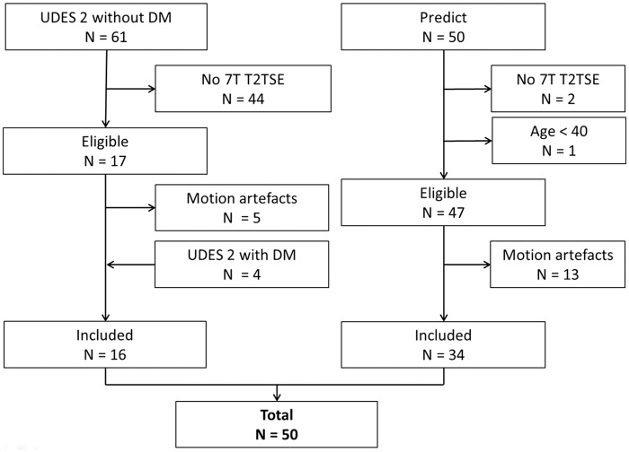
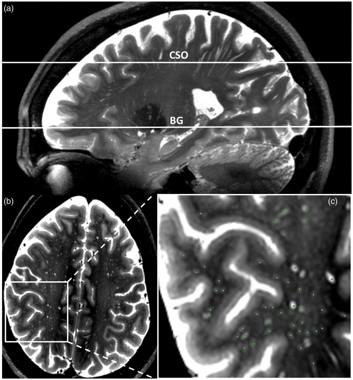
Cerebral perivascular spaces (PVS) are small physiological structures around blood vessels in the brain. MRI visible PVS are associated with ageing and cerebral small vessel disease (SVD). 7 Tesla (7T) MRI improves PVS detection. We investigated the association of age, vascular risk factors, and imaging markers of SVD with PVS counts on 7 T MRI, in 50 persons aged ≥ 40. The average PVS count ± SD in the right hemisphere was 17 ± 6 in the basal ganglia and 71 ± 28 in the semioval centre. We observed no relation between age or vascular risk factors and PVS counts. The presence of microbleeds was related to more PVS in the basal ganglia (standardized beta 0.32; p = 0.04) and semioval centre (standardized beta 0.39; p = 0.01), and white matter hyperintensity volume to more PVS in the basal ganglia (standardized beta 0.41; p = 0.02). We conclude that PVS counts on 7T MRI are high and are related SVD markers, but not to age and vascular risk factors. This latter finding may indicate that due to the high sensitivity of 7T MRI, the correlation of PVS counts with age or vascular risk factors may be attenuated by the detection of "normal", non-pathological PVS.
Keywords: Cerebrovascular disease; MRI; aging; brain imaging; microcirculation; physiology.
© The Author(s) 2016.
Figures
References
-
- Wang P, Olbricht WL. Fluid mechanics in the perivascular space. J Theor Biol 2011; 274: 52–57. - PubMed
-
- Schley D, Carare-Nnadi R, Please CP, et al. Mechanisms to explain the reverse perivascular transport of solutes out of the brain. J Theor Biol 2006; 238: 962–974. - PubMed
-
- Wuerfel J, Haertle M, Waiczies H, et al. Perivascular spaces—MRI marker of inflammatory activity in the brain? Brain 2008; 131: 2332–2340. - PubMed
-
- Bouvy WH, Biessels GJ, Kuijf HJ, et al. Visualization of perivascular spaces and perforating arteries with 7 T magnetic resonance imaging. Invest Radiol 2014; 49: 307–313. - PubMed
Publication types
MeSH terms
Grants and funding
LinkOut - more resources
Full Text Sources
Other Literature Sources
Medical
Research Materials
