. 2016 Jun:82:64-71.
doi: 10.1016/j.yhbeh.2016.04.010. Epub 2016 May 4.

Early life stress accelerates behavioral and neural maturation of the hippocampus in male mice

Affiliations

Early life stress accelerates behavioral and neural maturation of the hippocampus in male mice

K Bath et al. Horm Behav. 2016 Jun.

Abstract

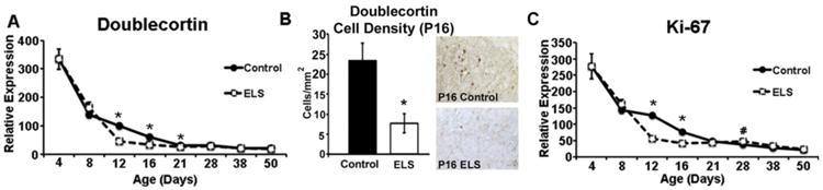
Early life stress (ELS) increases the risk for later cognitive and emotional dysfunction. ELS is known to truncate neural development through effects on suppressing cell birth, increasing cell death, and altering neuronal morphology, effects that have been associated with behavioral profiles indicative of precocious maturation. However, how earlier silencing of growth drives accelerated behavioral maturation has remained puzzling. Here, we test the novel hypothesis that, ELS drives a switch from growth to maturation to accelerate neural and behavioral development. To test this, we used a mouse model of ELS, fragmented maternal care, and a cross-sectional dense sampling approach focusing on hippocampus and measured effects of ELS on the ontogeny of behavioral development and biomarkers of neural maturation. Consistent with previous work, ELS was associated with an earlier developmental decline in expression of markers of cell proliferation (Ki-67) and differentiation (doublecortin). However, ELS also led to a precocious arrival of Parvalbumin-positive cells, led to an earlier switch in NMDA receptor subunit expression (marker of synaptic maturity), and was associated with an earlier rise in myelin basic protein expression (key component of the myelin sheath). In addition, in a contextual fear-conditioning task, ELS accelerated the timed developmental suppression of contextual fear. Together, these data provide support for the hypothesis that ELS serves to switch neurodevelopment from processes of growth to maturation and promotes accelerated development of some forms of emotional learning.


Conflict of interest statement

The authors declare no competing interest.

Figures

Fig. 1
Fig. 1
Experimental Design. We collected a wide variety of biochemical, genetic, histological, and behavioral measures across early development. Here we plot the timeline of our experimental manipulations, with bedding restriction occurring between P4 and P11. Both during and following this manipulation, we sampled from multiple separate litters of mice to collect basal serum corticosterone levels (Corticosterone), measures of early motor development (Motor Behavior), mRNA to assess gene expression (Gene expression), fixed brain tissue for immunohistochemistry (IHC), and carried out contextual fear conditioning in separate sets of mice (conditioning) and testing for fear expression 24 h following conditioning (Freezing Behavior). For all manipulations, individual animals contributed to only a single measure with unique cohorts of animals being used for the collection of each time point.
Fig. 2
Fig. 2
ELS significantly alters somatic and HPA development. A) Plots of the mean basal serum corticosterone levels sampled across early development. B) Plots of the mean weight for control (closed) and stressed (open) mice. C) Mean hang duration of mice on the inverted wire hang task at 17 days of age (P17). D) Plots of average distance traveled in the open field at 12 days of age (P12) and 21 days of age (P21). For all plots, error depicts the standard error of the mean. *p < 0.05.
Fig. 3
Fig. 3
ELS accelerates the developmental suppression of contextual fear responding. Mean percent duration freezing (±SEM) for the 5-minute context test (24 hours post conditioning). Each time point represents a different cohort of mice, sampled from multiple litters. A) Control mice (solid) showed a significant reduction in freezing at 29 days of age, compared with other developmental time points. B) In ELS mice (unfilled), a significant reduction in freezing was observed at 22 days of age compared with mice tested at all other developmental time points. C) Visual comparison of lines of best fit tracking developmental change in freezing behavior for control (solid) and ELS (dashed) mice, show a shift in the trough for freezing behavioral over development, with ELS mice decreasing earlier that control mice. Two-way ANOVA was used to assess overall effects of trial and between groups effects. Within group comparisons (n = 7 to 18 mice per group) were computed using one-way ANOVA with post hoc (Tukey's LSD) comparisons to test for effects of age. *p < 0.05.
Fig. 4
Fig. 4
ELS accelerates the developmental expression of markers of neural and circuit maturation. A) Realtime qPCR (RT-qPCR) tracking developmental change in the mean expression (±SEM) of parvalbumin (PV) normalized to beta-actin for control (solid) and ELS (dashed) samples (n = 5 per group). B) Quantification of parvalbumin-positive cell density in the dentate gyrus of the hippocampus across development, along with representative photomicrographs of PV-labeling in 21 day old control and ELS brains. C) Plot of the mean ratio of NR2a/NR2b mRNA expression across development for control and ELS hippocampus. D) Plot of relative mRNA expression (±SEM) of myelin basic protein (MBP) over development. Two-way ANOVA was used to assess overall effects of age and treatment × age interactions. Developmental shift in the ration of NR2a:NR2b was assessed using a single sample t-test (relative to 1). # indicates first point in which the ratio of NR2a:NR2b is significantly above 1 (equal ratio). Follow-up comparisons of treatment effects within age were carried out with planned un-paired Student's t-tests. *p < 0.05. #p < 0.05.
Fig. 5
Fig. 5
ELS accelerates the developmental decline in expression of markers of growth. A) RT-qPCR to assess doublecortin (DCX) mRNA levels across development in hippocampus of control and ELS mice. B) Quantification of DCX-positive cell density in subiculum of 16 day old control and ELS mice, along with representative photomicrographs of DCX-labeling. C) Plots of the developmental expression of Ki-67, a marker expressed by proliferating cells. All data are presented as mean (±SEM). RT-qPCR were normalized to beta-actin and derived from n = 5 samples per group. Two-way ANOVA was used to assess overall effects of age and treatment × age interactions. Follow up comparisons of treatment effects within age were carried out with planned un-paired Student's t-tests. *p < 0.05.
Fig. 6
Fig. 6
ELS leads to suppressed expression of GR and MR in the hippocampus. RT-qPCR to assess gene expression in hippocampus across development. Mean expression (±SEM) normalized to beta-actin is presented for control (solid) and ELS (dashed) samples (n = 5 per group). A) developmental expression of glucocorticoid receptor (GR) and B) developmental expression of mineralocorticoid receptor (MR). Two-way ANOVA was used to assess overall effects of age and treatment × age interactions. Follow up comparisons of treatment effects within age were carried out with planned un-paired Student's t-tests. *p < 0.05.

References

    1. Akers KG, Arruda-Carvalho M, Josselyn SA, Frankland PW. Ontogeny of contextual fear memory formation, specificity, and persistence in mice. Learn Mem. 2012;19:598–604. - PubMed
    1. Allsworth JE, Weitzen S, Boardman LA. Early age at menarche and allostatic load: data from the Third National Health and Nutrition Examination Survey. Ann Epidemiol. 2005;15:438–444. - PubMed
    1. Anda RF, Felitti VJ, Bremner JD, Walker JD, Whitfield C, Perry BD, Dube SR, Giles WH. The enduring effects of abuse and related adverse experiences in childhood. A convergence of evidence from neurobiology and epidemiology. Eur Arch Psychiatry Clin Neurosci. 2006;256:174–186. - PMC - PubMed
    1. Andersen SL, Teicher MH. Delayed effects of early stress on hippocampal development. Neuropsychopharmacology. 2004;29:1988–1993. - PubMed
    1. Avishai-Eliner S, Hatalski CG, Tabachnik E, Eghbal-Ahmadi M, Baram TZ. Differential regulation of glucocorticoid receptor messenger RNA (GR-mRNA) by maternal deprivation in immature rat hypothalamus and limbic regions. Brain Res Dev Brain Res. 1999;114:265–268. - PMC - PubMed