

Review
doi: 10.1016/j.ymeth.2016.04.032.
Epub 2016 May 4.
In vitro selection and amplification protocols for isolation of aptameric sensors for small molecules
Affiliations
- PMID: 27155227
- PMCID: PMC4981533
- DOI: 10.1016/j.ymeth.2016.04.032
Item in Clipboard
Review
In vitro selection and amplification protocols for isolation of aptameric sensors for small molecules
Methods.
.
Abstract
We recently optimized a procedure that directly yields aptameric sensors for small molecules in so-called structure-switching format. The protocol has a high success rate, short time, and is sufficiently simple to be readily implemented in a non-specialist laboratory. We provide a stepwise guide to this selection protocol.
Keywords: Aptamer; Biosensor; SELEX; Small molecules; Structure-switching.
Copyright © 2016 Elsevier Inc. All rights reserved.
Figures
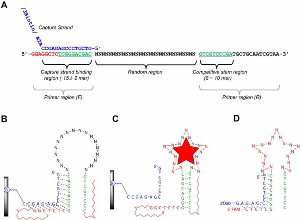
An example of SELEX library from Yang et al. [5]. (A) Library structure. This figure shows the relationship between a capture binding region and competitive stem region (green letters in both primer regions) which can induce the structural-switching upon target binding for the partitioning step. (B) An illustration of 2D structure of library binding on the streptavidin coated agarose column. A library element is attached to a column via a capture strand (blue letter in Fig. 1A). The capture strand is attached to an agarose-streptavidin column via a biotinylated oligonucleotide. “B” indicates biotinylation. (C) A library element partitioning mechanism. Upon binding the target, appropriate folding in the random region consequently induces the structural changes, which subsequently leads to dissociation from the capture stand. (D) An example of sensor design. After completion of SELEX, the final sensor is derived from trimming the flanking region of library element.
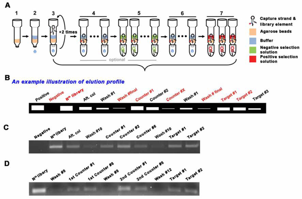
Schematic drawing for selection procedure. (A) (1) Agarose bead addition, (2) Wash the beads with SELEX buffer for equilbration, (3) Addition of the mixture of library and capture. After adding the mixture, the eluent is collected and re-applied into the column. This is to maximize the binding of the library-capture hybrid to the column. (4) Wash the column with the SELEX buffer ~10 times to remove uncaptured library elements. The number of washes can be adjusted based on the desired selection stringency. (5) Negative/Counter selection (optional). This elution step is to remove the less specific library elements that can bind to molecules which may compete with the desired target. (6) Additional wash - to remove residual solution from the negative selection, if applied. (7) Positive selection. During selection, several fractions need to be collected to monitor the elution profile. The choice of eluents to collect may be flexible, but the red labelled fractions should be collected, especially, the Nth library solution, the final buffer wash solution and the target solutions (#1 ~ #2). (B) An example illustration of elution profile. The collected eluents are compared though small scale PCR. The initial library templated PCR product can be utilized as a positive control for the PCR as well as the molecular weight marker. The commercial DNA ladder is used only when it is needed. The gel image of elution profile when one type of counter target is introduced (C), when two counter targets are applied (D).
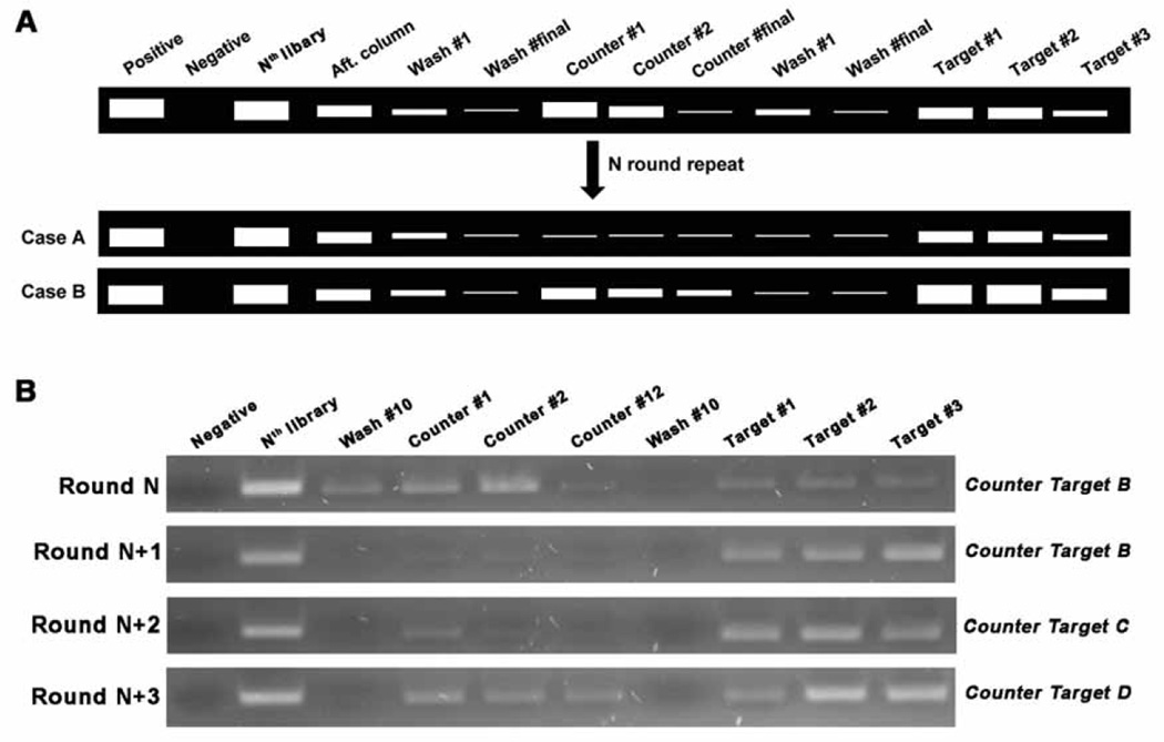
The stop point of negative/counter selection. (A) After introducing negative selections, more rounds are carried out until the elution bands are dramatically decreased (Case A) or the target elution bands are brighter than the counter target elution bands (Case B). (B) Gel image example from “Target A” SELEX. At Round N (e.g. Round 9), ‘B’ was introduced as the counter target molecule, which is one of the analogs of target A, and showed similar elution bands in the counter and target lanes. At Round N+1, this counter target no longer bound to the library elements as observed by the lack of elution bands in the counter lanes. In the following round (Round N+2), we used another analog, ‘C,’ as the counter target, and observed a faint band in the counter target lanes of much weaker fluorescent intensity than in the target elution lanes. We then applied a third analog, ‘D,’ as the counter target in the next round (Round N+3) and we again observed some bands of much weaker fluorescent intensity than when the target was used. For the next two rounds, ‘D’ was used as a counter target and a similar elution profile was observed to Round (N + 3). This prompted us to stop the SELEX process and begin cloning.
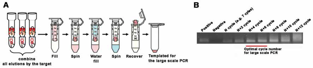
Large scale PCR. (A) Template preparation for the large scale PCR. Collect all target elutions, concentrate using a 3K MWCO centrifugal filter until it reaches 50 ~ 100 µL, then wash this concentrated solution with 400 ~ 500 µL of DNase/RNase-free water. This water washing is critical for high efficiency of large scale PCR. Use of all collected solution is recommended up until the ~5th round to allow amplification of all the oligonucleotide strands. After the 5th round, we recommend using only ~50% of template or less. The advantage of doing this is to keep a backup should the following rounds of SELEX fail, and after the 6th round, there are already multiple copies for many of the strands. (B) An example of small scale PCR (50 µL) for deciding the number of cycles before carrying out the large scale PCR (1 mL). Collect the PCR product after adding 2 cycles each time, and choose the number of cycles that give the highest yield, but not over-amplified. In this case, 11 ~ 13 cycles is recommended.
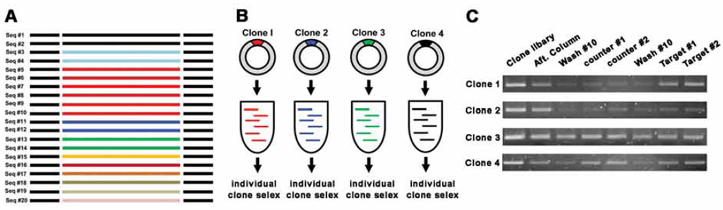
A schematic for clone SELEX. (A) After sequencing, count repeats and choose ~ 4 clones based on their abundance. (B) Large scale PCR and strand separation for the selected clones. Strands of one particular sequence is amplified through large scale PCR and library is prepared. (C) Representative cases of clone SELEX elution profile. The elution profile of Clone 1 is the best, clone 4 is second. Cases 2 and 3 are not recommended for adaptation into sensors.
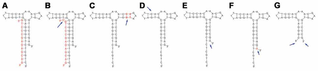
An example of sensor optimization. This is a steroid aptamer for deoxycorticosterone 21-glucoside [4]. Based on the original sensor (A), extended capture length (B), extended side stem (C), shortened side stem (D), shortened main stem length (E), or extended main stem base paring (F). (G) is the shortened sensor form for utilizing without the capture. Arrows indicate the modification sites.
Similar articles
-
Aptamers: The "evolution" of SELEX.Methods. 2016 Aug 15;106:21-8. doi: 10.1016/j.ymeth.2016.04.020. Epub 2016 Apr 19. Methods. 2016. PMID: 27109056 Review.
-
Strategies for Creating Structure-Switching Aptamers.ACS Sens. 2018 Sep 28;3(9):1611-1615. doi: 10.1021/acssensors.8b00516. Epub 2018 Aug 29. ACS Sens. 2018. PMID: 30156834
-
Exploration of structure-switching in the design of aptamer biosensors.Adv Biochem Eng Biotechnol. 2014;140:69-92. doi: 10.1007/10_2013_223. Adv Biochem Eng Biotechnol. 2014. PMID: 23851586
-
Advances in aptamer screening and small molecule aptasensors.Adv Biochem Eng Biotechnol. 2014;140:29-67. doi: 10.1007/10_2013_225. Adv Biochem Eng Biotechnol. 2014. PMID: 23851587 Review.
-
Electrochemical nanomaterial-based nucleic acid aptasensors.Anal Bioanal Chem. 2012 Apr;402(10):3103-14. doi: 10.1007/s00216-012-5769-1. Epub 2012 Feb 16. Anal Bioanal Chem. 2012. PMID: 22349328 Review.
Cited by
-
Effects of storage conditions on the performance of an electrochemical aptamer-based sensor.Sens Diagn. 2024 May 14;3(6):1044-1050. doi: 10.1039/d4sd00066h. eCollection 2024 Jun 13. Sens Diagn. 2024. PMID: 38882472 Free PMC article.
-
Real-Time, In Vivo Molecular Monitoring Using Electrochemical Aptamer Based Sensors: Opportunities and Challenges.ACS Sens. 2022 Oct 28;7(10):2823-2832. doi: 10.1021/acssensors.2c01428. Epub 2022 Oct 7. ACS Sens. 2022. PMID: 36205360 Free PMC article. Review.
-
Highly selective DNA aptamer sensor for intracellular detection of coenzyme A.Chem Sci. 2025 Mar 28;16(18):8023-8029. doi: 10.1039/d5sc00332f. eCollection 2025 May 7. Chem Sci. 2025. PMID: 40206557 Free PMC article.
-
Tandem metabolic reaction-based sensors unlock in vivo metabolomics.Proc Natl Acad Sci U S A. 2025 Mar 4;122(9):e2425526122. doi: 10.1073/pnas.2425526122. Epub 2025 Feb 27. Proc Natl Acad Sci U S A. 2025. PMID: 40014569 Free PMC article.
-
A massively parallel screening platform for converting aptamers into molecular switches.Nat Commun. 2023 Apr 24;14(1):2336. doi: 10.1038/s41467-023-38105-4. Nat Commun. 2023. PMID: 37095144 Free PMC article.
References
-
- Darmostuk M, Rimpelova S, Gbelcova H, Ruml T. Biotechnol. Adv. 2015;33:1141–1161. http://dx.doi.org/10.1016/j.biotechadv.2015.02.008. - DOI - PubMed
-
- Sun H, Zu Y. Molecules. 2015;20:11959–11980. http://dx.doi.org/10.3390/molecules200711959. - DOI - PMC - PubMed
-
- Blind M, Blank M. Mol. Ther. Nucleic Acids. 2015:e233. http://dx.doi.org/10.1038/mtna.2014.74. - DOI - PMC - PubMed
-
- Yang K-A, Pei R, Stefanovic D, Stojanovic MN. J. Am. Chem. Soc. 2012;134:1642–1647. http://dx.doi.org/10.1021/ja2084256. - DOI - PubMed
-
- Yang K-A, Barbu MB, Halim M, Pallavi P, Kim B, Kolpashchikov DM, Pecic S, Taylor S, Worgall TS, Stojanovic MN. Nat. Chem. 2014;6:1003–1008. http://dx.doi.org/10.1038/nchem.2058. - DOI - PMC - PubMed
Publication types
MeSH terms
Substances
Grants and funding
LinkOut - more resources
Full Text Sources
Other Literature Sources
