

Comment
doi: 10.1016/j.jid.2016.04.019.
Epub 2016 May 5.
Nucleophagy: A New Look at Past Observations
Affiliations
- PMID: 27155459
- PMCID: PMC7383959
- DOI: 10.1016/j.jid.2016.04.019
Item in Clipboard
Comment
Nucleophagy: A New Look at Past Observations
J Invest Dermatol.
2016 Jul.
Abstract
Keratinization of the stratum corneum involves a highly choreographed sequence of events in which granular cells lose their nuclei and become desiccated corneocytes. Akinduro et al. detail the molecular machinery underlying removal of the nucleus (nucleophagy) during the final stages of keratinization. They provide evidence that nucleophagy is induced when the keratinocytes differentiate and that failure in the initiation of nucleophagy is associated with parakeratosis.
Copyright © 2016 The Authors. Published by Elsevier Inc. All rights reserved.
Figures
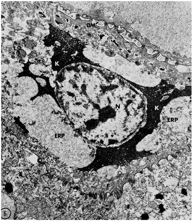
A keratinocyte in an advanced stage of transformation from a granular
cell to a corneocyte. Lysosomes/autophagosomes (L) are present in the cytoplasm.
Filaggrin (K) is spread throughout the cytoplasm intermingling with keratin
filaments (F). The thickened, modified corneocyte envelope (PM) is tightly
attached to the lower granular cell. The nucleus (N), last of the recognizable
organelles, shows signs of degradation. Reprinted from ©Lavker and Matoltsy, 1970, J Cell Biology,
44:501–512
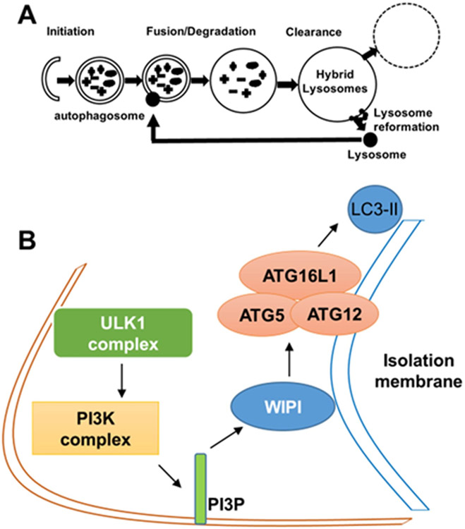
Autophagy and its initiation. (A) A schematic representation of the
stages of autophagy. (B) A schematic representation of the initial steps in
autophagy involving the formation of an isolation membrane. In this process,
unc-51 like autophagy activating kinase 1 (ULK1) complex activates the
phosphatidylinositol-3-kinase (PI3K) complex (VPS34/Beclin1/ATG14L).
VPS34-derived PI3P recruits double FYVE-containing protein 1 (DFCP1/ZFYVE1) and
WD repeat domain phosphoinositide interacting 1 (WIPI1) to the outer membrane of
autophagosomes, which causes the association of the ATG5/ATG12 conjugate with
ATG16L1. The ATG5/ATG12/ATG16L1 complex then adds phosphatidylethanolamine group
(PE) to the C-terminus of the microtubule associated protein 1 light chain 3
(LC3) protein promoting the elongation of the isolation membrane and
autophagosome formation.
Comment on
-
Constitutive Autophagy and Nucleophagy during Epidermal Differentiation.J Invest Dermatol. 2016 Jul;136(7):1460-1470. doi: 10.1016/j.jid.2016.03.016. Epub 2016 Mar 25. J Invest Dermatol. 2016. PMID: 27021405
References
-
- Candi E, Schmidt R, Melino G (2005) The cornified envelope: a model of cell death in the skin. Nat Rev Mol Cell Biol 6:328–40. - PubMed
-
- Chen Y, Yu L (2013) Autophagic lysosome reformation. Exp Cell Res 319:142–6. - PubMed
-
- Douroudis K, Kingo K, Traks T, Reimann E, Raud K, Ratsep R, et al. (2012) Polymorphisms in the ATG16L1 gene are associated with psoriasis vulgaris. Acta Derm Venereol 92:85–7. - PubMed
Publication types
MeSH terms
Grants and funding
LinkOut - more resources
Full Text Sources
Other Literature Sources
Miscellaneous
