Anatomic study suggests that the morphology of the plantaris tendon may be related to Achilles tendonitis
- PMID: 27155667
- PMCID: PMC5309290
- DOI: 10.1007/s00276-016-1682-1
Anatomic study suggests that the morphology of the plantaris tendon may be related to Achilles tendonitis
Abstract
Purpose: Achilles tendinopathy is a significant clinical lower limb issue observed in recent years. Neither the location nor the mechanism behind the pain has yet been sufficiently explained. Patients frequently experience pain on the medial side of the calcaneal tendon, and between 2 and 7 cm above the calcaneal tuberosity, which may suggests that the plantaris tendon plays an important role. The purpose of this study was to determine the anatomical relationships between the course of the plantaris tendon and the calcaneal tendon, as well as the type of insertion of the plantaris tendon.
Methods: The tests were carried out on 50 randomized lower limbs (23 left and 27 right) fixed in 10 % formalin solution.
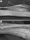
Results: Five insertion types of the plantaris tendon were identified in relation to the calcaneal tendon: four with their insertion on the calcaneal tuberosity (Types 1, 2, 3, 5), while the fifth (Type 4) had its insertion in the crural fascia. In addition, two variants of the course of the plantaris tendon were identified, the most common being termed Variant A, in which the tendon crosses the space between the gastrocnemius and the soleus muscles, thus reaching the medial crural region, and is located on the medial side of the calcaneal tendon (84 % cases). The course of the Variant B is similar to the course of the Variant A, but upon leaving the space located between the gastrocnemius and soleus muscle, it turned to the medial crural region and ran directly anterior to the calcaneal tendon (12 %). The plantaris muscle was found to be absent in two lower limbs (4 %). The most frequent insertion type of the plantaris tendon into the calcaneal tuberosity is fan-shaped, occurring on the medial side of the Achilles tendon (Type 1-44 % cases).
Conclusion: The course of the plantaris tendon and its mobility range in relation to the calcaneal tendon may be likely to affect the occurrence of pains in the lower medial part of the leg (Achilles tendinopathy).
Keywords: Calcaneal tendon; Mid-portion Achilles tendinopathy; Plantaris muscle; Plantaris tendon.
Conflict of interest statement
The authors declare that they have no conflict of interest.
Figures


Similar articles
-
The plantaris tendon and a potential role in mid-portion Achilles tendinopathy: an observational anatomical study.J Anat. 2011 Mar;218(3):336-41. doi: 10.1111/j.1469-7580.2011.01335.x. J Anat. 2011. PMID: 21323916 Free PMC article.
-
A highly complex variant of the plantaris tendon insertion and its potential clinical relevance.Anat Sci Int. 2020 Sep;95(4):553-558. doi: 10.1007/s12565-020-00540-4. Epub 2020 Apr 4. Anat Sci Int. 2020. PMID: 32248353 Free PMC article.
-
The Plantaris Muscle Tendon and Its Relationship with the Achilles Tendinopathy.Biomed Res Int. 2018 May 31;2018:9623579. doi: 10.1155/2018/9623579. eCollection 2018. Biomed Res Int. 2018. PMID: 29955614 Free PMC article.
-
The plantaris tendon: a narrative review focusing on anatomical features and clinical importance.Bone Joint J. 2016 Oct;98-B(10):1312-1319. doi: 10.1302/0301-620X.98B10.37939. Bone Joint J. 2016. PMID: 27694583 Review.
-
Cadaveric Anatomical Study of Sural Nerve: Where is The Safe Area for Endoscopic Gastrocnemius Recession?Open Orthop J. 2017 Sep 30;11:1094-1098. doi: 10.2174/1874325001711011094. eCollection 2017. Open Orthop J. 2017. PMID: 29152002 Free PMC article. Review.
Cited by
-
Is the plantaris muscle the most undefined human skeletal muscle?Anat Sci Int. 2021 Jun;96(3):471-477. doi: 10.1007/s12565-020-00586-4. Epub 2020 Nov 7. Anat Sci Int. 2021. PMID: 33159667 Free PMC article.
-
MRI of the Achilles tendon-A comprehensive pictorial review. Part one.Eur J Radiol Open. 2021 Mar 26;8:100342. doi: 10.1016/j.ejro.2021.100342. eCollection 2021. Eur J Radiol Open. 2021. PMID: 33850971 Free PMC article. Review.
-
[Anatomical and biomechanical characteristics of plantaris tendon and its application in ligament reconstruction].Zhongguo Xiu Fu Chong Jian Wai Ke Za Zhi. 2024 Feb 15;38(2):234-239. doi: 10.7507/1002-1892.202311032. Zhongguo Xiu Fu Chong Jian Wai Ke Za Zhi. 2024. PMID: 38385238 Free PMC article. Chinese.
-
Bicipital origin and the course of the plantaris muscle.Anat Cell Biol. 2021 Jun 30;54(2):289-291. doi: 10.5115/acb.21.086. Anat Cell Biol. 2021. PMID: 34053915 Free PMC article.
-
Impact of plantaris ligamentous tendon.Sci Rep. 2021 Feb 25;11(1):4550. doi: 10.1038/s41598-021-84186-w. Sci Rep. 2021. PMID: 33633305 Free PMC article.
References
-
- Anson BJ, McVay CB. Surgical Anatomy. Philadelphia: Saunders Company; 1971. pp. 1186–1189.
-
- Aragão JA, Reis FP, Guerra DR, Cabral RH. The occurrence of the plantaris muscle and it’s muscle-tendon relationship in adult human cadavers. Int J Morphol. 2010;28:255–258. doi: 10.4067/S0717-95022010000100037. - DOI
-
- Bergman RA, Afifi AK, Miyauchi R (2015) Illustrated Encyclopedia of Human Anatomic Variation: Opus I: Muscular System: Alphabetical Listing of Muscles. http://www.anatomyatlases.org/AnatomicVariants/MuscularSystem/Text/P/29P.... Accessed 13 Dec 2015
-
- Cummins EJ, Anson BJ, et al. The structure of the calcaneal tendon (of Achilles) in relation to orthopedic surgery with additional observation on the plantaris muscle. Surg Gynecol Obstet. 1946;83:107–116. - PubMed
MeSH terms
LinkOut - more resources
Full Text Sources
Other Literature Sources
Medical
Miscellaneous

