Review
. 2016 Jun;41(6):491-507.
doi: 10.1016/j.tibs.2016.04.002. Epub 2016 May 4.

RecA: Regulation and Mechanism of a Molecular Search Engine

Affiliations
Review

RecA: Regulation and Mechanism of a Molecular Search Engine

Jason C Bell et al. Trends Biochem Sci. 2016 Jun.

Erratum in

Abstract

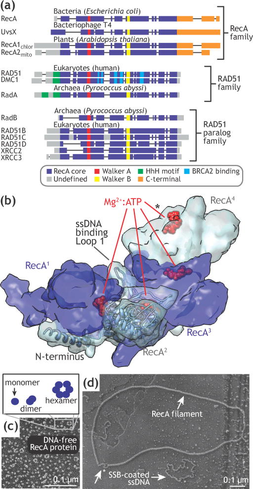
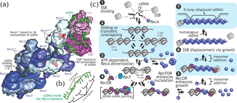
Homologous recombination maintains genomic integrity by repairing broken chromosomes. The broken chromosome is partially resected to produce single-stranded DNA (ssDNA) that is used to search for homologous double-stranded DNA (dsDNA). This homology driven 'search and rescue' is catalyzed by a class of DNA strand exchange proteins that are defined in relation to Escherichia coli RecA, which forms a filament on ssDNA. Here, we review the regulation of RecA filament assembly and the mechanism by which RecA quickly and efficiently searches for and identifies a unique homologous sequence among a vast excess of heterologous DNA. Given that RecA is the prototypic DNA strand exchange protein, its behavior affords insight into the actions of eukaryotic RAD51 orthologs and their regulators, BRCA2 and other tumor suppressors.

Keywords: Förster resonance energy transfer (FRET); RAD51; homology search; recombination; self-assembly; single molecule imaging; total internal reflection fluorescence (TIRF).


Figures

Figure 1
Figure 1. RecA conservation and structure
A) Conserved domains of the RecA, RAD51, and RAD51 paralog families. B) Segment of RecA filament showing the Mg:ATP binding site at the monomer-monomer interface. The asterisk indicates a half-site; for simplicity, only one of ssDNA binding loops per monomer is shown. Adapted from PDB 3CMW (RecA) [30]. C) Electron microscopy of DNA-free RecA showing heterogeneous oligomerization states of RecA. D) Electron microscopy of RecA filament-formation on SSB-coated circular ssDNA. Note the dramatic extension of the RecA filament relative to the compaction of the SSB-coated ssDNA. C) and D) adapted from [39].
Figure 2
Figure 2. Mechanism of RecA nucleation and growth on SSB-coated ssDNA
A) Structural model of twelve-monomers of RecA bound to 36 nucleotides of ssDNA adjacent to two tetramers of SSB bound to 140 nucleotides of ssDNA demonstrating the vast differences in site size and accessibility of the ssDNA bound to each protein. Adapted from PDB 3CMW (RecA) and PDB 1EYG (SSB) [30, 37]. Mg:ATP (red) is visible at the monomer-monomer interface for RecA7-RecA12. B) Single-stranded DNA within the filament is stretched into nucleotide triplets that can maintain Watson-Crick interactions during homologous pairing. C) 1, SSB binds rapidly to ssDNA, removing secondary structure that impedes RecA-mediated DNA strand exchange. 2, SSB kinetically blocks RecA filament formation. 3, Nucleation of RecA onto rare and transient microscopic gaps requires ATP-dependent dimerization, making nucleation infrequent. 4, RecOR binds to the C-terminal tails of SSB, microscopically altering the SSB-ssDNA complex, but not displacing it. This ‘stoichiometric remodeling’ creates microscopic gaps that are large enough and long-lived enough for RecA to stably bind, enhancing nucleation. Similarly, RecF further, in coordination with RecOR, enhances RecA nucleation at dsDNA-ssDNA junctions. 5, RecA filament growth through monomer addition is impeded by SSB, though less so than nucleation; however, in the presence of RecOR, RecA filament growth is stimulated ~3-fold. 6, The RecA filament grows monotonically and displaces SSB from ssDNA. Adapted from [24]. 7, The RecA-ssDNA filament catalyzes pairing and strand exchange with a homologous dsDNA molecule, resulting in an intermediate, three-stranded molecule called a D-loop (or ‘displacement loop’); SSB binds to the displaced strand to stabilize the D-loop (not shown).
Figure 3
Figure 3. Diffusion-driven mechanism of RecA-mediated homology search
A) Diagram of three particles demonstrating random walk diffusion represented on a two-dimensional plane. Adapted from reference [117]. B) Plot showing the mean squared displacement as a function of time for one-, two-, and three-dimensional diffusion compared to three-dimensional diffusion in a confined space without or with directed motion Adapted from [99, 118]. C) Cartoon depicting different modes by which proteins find their targets by sliding, hopping, jumping, intersegmental transfer, and intersegmental contact sampling [–72, 75]. Single-molecule methods used to measure RecA-mediated homology search: D) Total internal reflection fluorescence (TIRF) microscopy used to visualize ssDNA-RecA filaments pairing with λ phage DNA [86]. E) DNA micromanipulation experiments demonstrating RecA pairing efficiency increases as the DNA is allowed to adopt three-dimensional, random-coil configurations [86]. F) Single-molecule Förster Resonance Energy Transfer (FRET) experiments used to demonstrate microscale sliding of RecA filaments [89].
Figure 4
Figure 4. Reduction in complexity through microhomology sampling
A) “Dual-molecule” method simultaneously using magnetic and optical traps to measure filament-DNA contacts formed on stretched or supercoiled DNA [90]. B) Molecular modeling and simulation of dsDNA interactions with the RecA-ssDNA nucleoprotein filament during initial contact, binding to the secondary DNA binding site (site II) and homology sampling. Adapted from [91]. C) Molecular model of a RecA pairing intermediate [91, 92]. Labeled amino acids indicate positively charged residues that constitute the secondary DNA binding site that binds to and stabilizes the incoming homologous dsDNA. D) Schematic and E) representative data from a single-molecule ‘DNA curtain’ experiment measuring RecA-mediated DNA ‘microhomology sampling’ between a long RecA-ssDNA filament and short, fluorescently-labeled dsDNA molecules [94]. F) Molecular modeling (for simplicity, only two strands are shown) of the transient kinetic intermediates formed during the initial microhomology sampling (8 base pairs or less) and subsequent DNA pairing reaction (9 base pairs or greater), resulting in the post-synaptic, Rad51/RecA-Stretched DNA intermediate that is stretched into segments of three base pairs containing normal Watson-Crick spacing, interspersed with 7–8 Å gaps formed by intercalating hydrophobic residues (e.g., Met164 and Ile99). Adapted from [95].

References

    1. Lin Z, et al. Origins and evolution of the recA/RAD51 gene family: evidence for ancient gene duplication and endosymbiotic gene transfer. Proc Natl Acad Sci U S A. 2006;103:10328–10333. - PMC - PubMed
    1. Rocha EP, et al. Comparative and evolutionary analysis of the bacterial homologous recombination systems. PLoS Genet. 2005;1:e15. - PMC - PubMed
    1. Wu D, et al. Stalking the fourth domain in metagenomic data: searching for, discovering, and interpreting novel, deep branches in marker gene phylogenetic trees. PLoS ONE. 2011;6:e18011. - PMC - PubMed
    1. Chintapalli SV, et al. Reevaluation of the evolutionary events within recA/RAD51 phylogeny. BMC Genomics. 2013;14:240. - PMC - PubMed
    1. Bianco PR, et al. DNA strand exchange proteins: A biochemical and physical comparison. Front Biosci. 1998;3:D570–603. - PubMed

MeSH terms