. 2016 May 19;165(5):1106-1119.
doi: 10.1016/j.cell.2016.04.015. Epub 2016 May 5.

Bacterial Outer Membrane Vesicles Mediate Cytosolic Localization of LPS and Caspase-11 Activation

Affiliations

Bacterial Outer Membrane Vesicles Mediate Cytosolic Localization of LPS and Caspase-11 Activation

Sivapriya Kailasan Vanaja et al. Cell. .

Abstract

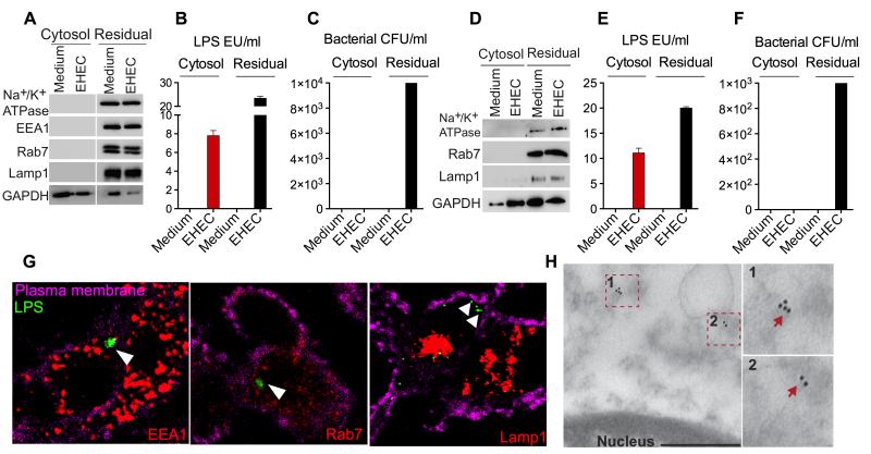
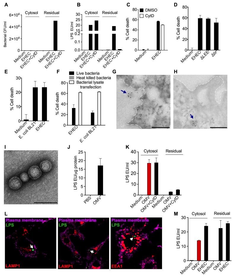
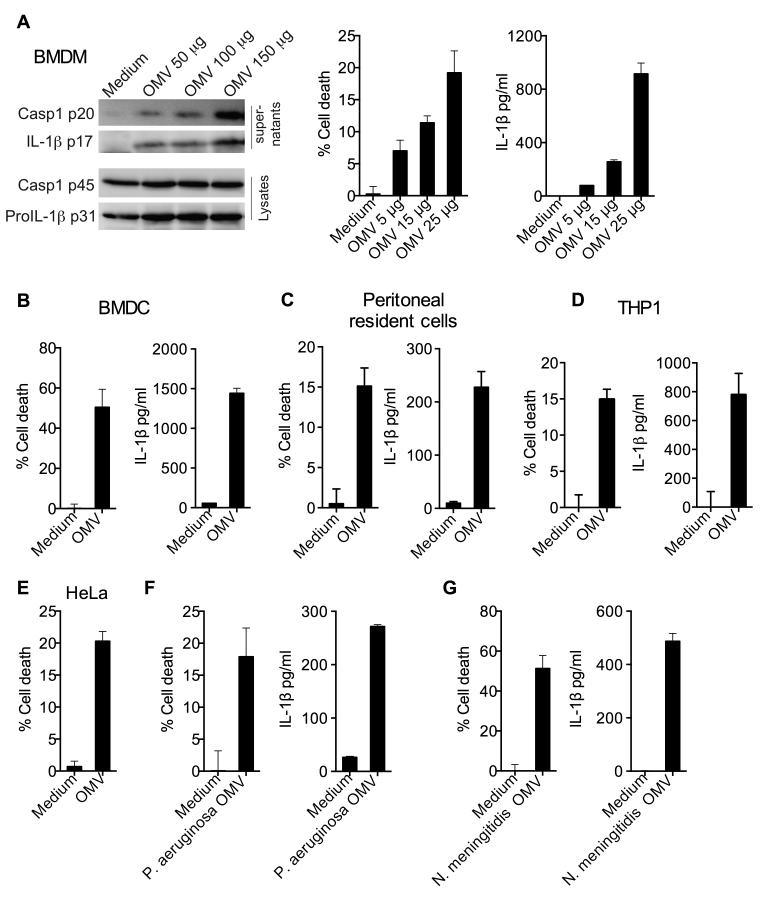
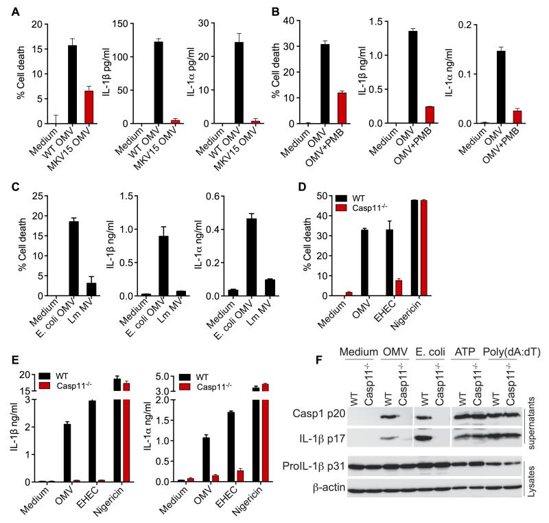
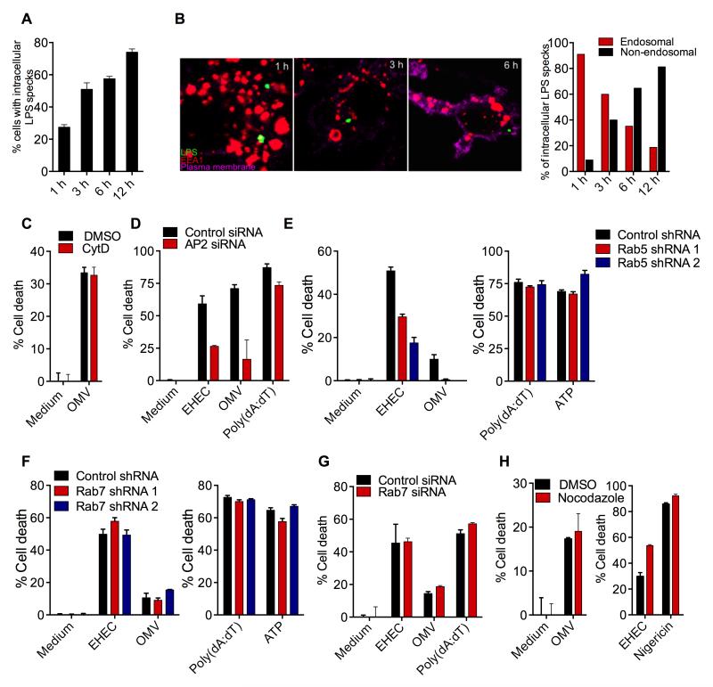
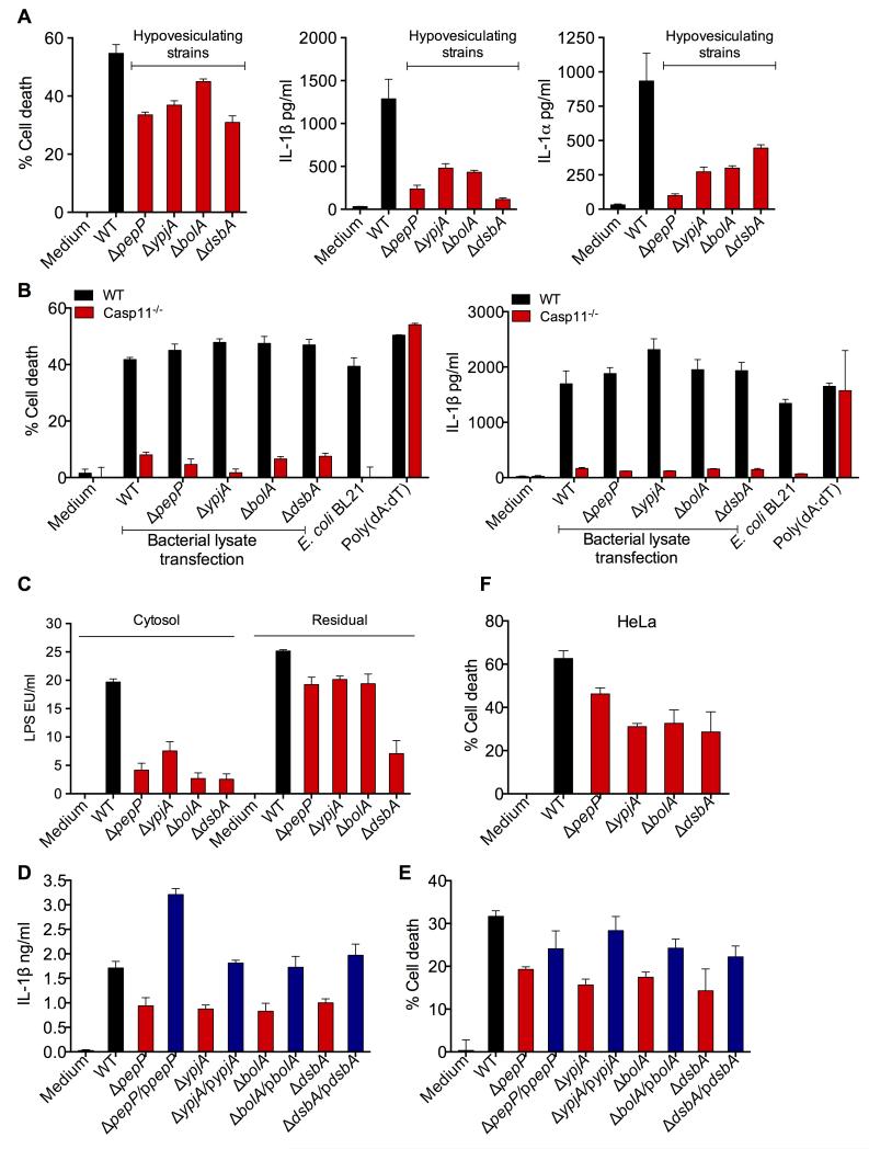
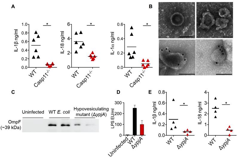
Sensing of lipopolysaccharide (LPS) in the cytosol triggers caspase-11 activation and is central to host defense against Gram-negative bacterial infections and to the pathogenesis of sepsis. Most Gram-negative bacteria that activate caspase-11, however, are not cytosolic, and the mechanism by which LPS from these bacteria gains access to caspase-11 in the cytosol remains elusive. Here, we identify outer membrane vesicles (OMVs) produced by Gram-negative bacteria as a vehicle that delivers LPS into the cytosol triggering caspase-11-dependent effector responses in vitro and in vivo. OMVs are internalized via endocytosis, and LPS is released into the cytosol from early endosomes. The use of hypovesiculating bacterial mutants, compromised in their ability to generate OMVs, reveals the importance of OMVs in mediating the cytosolic localization of LPS. Collectively, these findings demonstrate a critical role for OMVs in enabling the cytosolic entry of LPS and, consequently, caspase-11 activation during Gram-negative bacterial infections.


Figures

Figure 1
Figure 1. LPS Gains Access to the Cytosol During Extracellular Gram-Negative Bacterial Infections
(A-C) Immunoblots for Na+/K+ ATPase, EEA1, Rab7, LAMP1 and GAPDH (A), LAL assay for LPS (EU, endotoxin units; B), and agar plating for bacterial counts (C) in the cytosolic and residual fractions of uninfected BMDM or BMDM infected with EHEC for 4 h at an MOI of 10 obtained by digitonin fractionation. (D-F) Immunoblots for Na+/K+ ATPase, Rab7, LAMP1 and GAPDH (D), LAL assay for LPS (E) and agar plating for bacterial counts (F) in the cytosolic and residual fractions of uninfected or EHEC-infected BMDM obtained by Dounce homogenization in hypotonic buffer. (G) Confocal microscopy of BMDM infected with EHEC at an MOI of 10 for 4 h. LPS was visualized with an anti-LPS antibody (green), EEA1, Rab7 and LAMP1 with the respective antibodies (red) and plasma membrane with cholera toxin B (magenta). (H) Transmission electron microscopy (TEM) of BMDM infected with EHEC for 4 h at an MOI of 10 following immunogold labeling with an anti-LPS antibody. Scale bar = 0.5 μM. Data are presented as mean±SEM from one experiment representative of three experiments for A-C and two experiments for D-H. See also Figure S1.
Figure 2
Figure 2. Bacterial OMV Deliver LPS into the Cytosol
(A-B) Bacterial count (A) and LPS quantity (B) as determined by agar plating and LAL assay, respectively, in the cytosolic and residual fractions from uninfected or EHEC-infected BMDM pretreated with DMSO or 2 μM cytochalasin D (cytD) 45 min prior to infection. (C) Cell death in uninfected or EHEC-infected BMDM pretreated with DMSO or 2 μM cytochalasin D (cytD) 45 min prior to infection as determined by Cell titer glo assay at 16 h post-infection (p.i). (D-E) Cell death in BMDM following infection with the indicated bacterial strains as determined by LDH assay at 16 h p.i. (F) Cell death in BMDM following stimulation with live or heat killed bacteria or transfection with bacterial lysate as determined by LDH assay at 16 h p.i. (G-H) Transmission electron microscopy (TEM) of BMDM infected with EHEC for 4 h at an MOI of 10 following immunogold labeling with an anti-LPS antibody. Plasma membrane is depicted by dotted lines. Scale bar = 0.5 μM. (I-J) Negative staining TEM of purified OMV (I) and LAL assay for LPS in purified OMV (J). Scale bar = 0.1 μM. (K) LPS levels in the cytosolic and residual fractions of BMDM treated with 50 μg OMV for 4 h as assessed by LAL assay. (L) Confocal images of BMDM treated with 25 μg of OMV for 4 h. Cells were stained for LPS, EEA1 or LAMP1 with corresponding antibodies and for plasma membrane with cholera toxin B. Arrows indicate LPS staining. (M) LPS levels in the cytosolic and residual fractions of HeLa cells treated with 200 μg OMV or EHEC at an MOI of 300 for 6 h as assessed by LAL assay. Data are presented as mean±SEM of one experiment representative of two experiments. See also Figure S2.
Figure 3
Figure 3. Bacterial OMV Activate Cell Death and IL-1 Responses
(A) Immunoblots for indicated proteins, LDH assay for cell death and ELISA for IL-1β in wild-type BMDM treated with indicated doses of OMV for 16 h. (B-G) Cell death and/or IL-1β secretion by indicated cell types treated with 25 μg of indicated OMV for 16 h. Data are presented as mean±SEM of one experiment representative of two experiments.
Figure 4
Figure 4. Activation of Cell Death and IL-1 by OMV is Dependent on LPS and Caspase-11
(A-C) Cell death and IL-1β and IL-1α secretion by BMDM treated with OMV from wild-type E. coli or MKV15 strain, which lacks a functional hexa-acylated lipid A (A) or OMV pretreated with 10 μg/ml polymyxin B (PMB) (B) or membrane vesicles from L. monocytogenes (Lm MV) (C). (D-E) Cell death and secretion of IL-1β and IL-1α by wild-type or caspase-11−/− BMDM stimulated as indicated for 16 h. (F) Cleaved caspase-1 p20 and IL-1β p17 in the supernatants and proIL-1β and β-actin in the lysates of wild-type or caspase-11−/− BMDM stimulated as indicated for 16 h. Data are presented as mean±SEM of one experiment representative of two experiments for A-C and F and three experiments for D-E. See also Figure S3.
Figure 5
Figure 5. Bacterial OMV Attain Intracellular Access through Endocytosis
(A-B) BMDM were treated with OMV for the indicated time points and were stained with antibodies against LPS and EEA1 as well as cholera toxin B for plasma membrane. Confocal microscopy-based quantification of % of cells with intracellular LPS (A) or % of intracellular LPS in the endosomal vs nonendosomal compartments (B). Quantification was done by counting 50 fields containing ~10 cells each. (C and H) Cell death in BMDM pretreated with 2 μM cytochalasin D (cytD) or 10 μM nocodazole 45 min prior to stimulation with the indicated treatments. (D) Cell death in immortalized BMDM (iBMDM) transfected with control or AP2 siRNA and treated as indicated. (E-F) Cell death in iBMDM expressing control, Rab5A (E), or Rab7 (F) shRNAs in response to indicated stimulations. (G) Cell death in iBMDM transfected with control or Rab7 siRNA and treated as indicated. Data are presented as mean±SEM of one experiment representative of two experiments. See also Figure S4.
Figure 6
Figure 6. OMV Production is Essential for E. coli-induced Caspase-11 Activation
(A) Cell death and secretion of IL-1β and IL-1α by unprimed BMDM stimulated with wild-type or indicated hypovesiculating strains of E. coli at an MOI of 25 for 16 h. (B) Cell death and secretion of IL-1β by wild-type or caspase-11−/− BMDM transfected with lysates from wild-type or indicated hypovesiculating strains of E. coli or stimulated with E. coli or poly(dA:dT) for 16 h. (C) LPS levels, as determined by LAL assay, in the cytosol and residual fractions of BMDM infected with wild-type or indicated hypovesiculating strains of E. coli for 4 h. (D-E) Secretion of IL-1β and cell death in BMDM infected with wild-type or indicated hypovesiculating strains or their corresponding complements expressing the deleted gene on a plasmid (blue bars) at an MOI of 25 for 16 h. (F) Cell death in HeLa cells stimulated with wild-type or indicated hypovesiculating strains of E. coli at an MOI of 300 for 16 h. Data are presented as mean±SEM of one experiment representative of three experiments for AB and two experiments for C-F. See also Figure S5.
Figure 7
Figure 7. OMV are Essential for the Activation of Cytosolic LPS Sensing in vivo
(A) IL-1β, IL-18, and IL-1α levels in the plasma of wild-type and caspase-11−/− mice injected i.p. with 100 μg of OMV. Mice were first primed with 200 μg poly(I:C) for 6 h (i.p.). Cytokine levels were assessed 6 h post OMV injection (n=6). (B) Negative staining (top row) and LPS immunogold (bottom row) TEM for OMV in the peritoneal lavages from wild-type mice infected i.p with 109 CFU of E. coli for 12 h (images were from two different mice). Scale bar = 0.1 μM. (C-D) Immunoblot for OmpF (C) and LAL assay for LPS (D) in OMV isolated from the peritoneal lavages of mice infected with 109 CFU of wild-type or ΔypjA strain of E. coli for 12 h. Each lane represents one mouse. (E) IL-1β and IL-18 levels in the plasma of wild-type mice infected with 109 CFU of wild-type or ΔypjA strain of E. coli for 12 h (n=4). Data are presented as mean±SEM of one experiment representative of two experiments. See also Figure S6.

Comment in

References

    1. Acevedo R, Fernández S, Zayas C, Acosta A, Sarmiento ME, Ferro VA, Rosenqvist E, Campa C, Cardoso D, Garcia L, et al. Bacterial outer membrane vesicles and vaccine applications. Front. Immunol. 2014;5:121. - PMC - PubMed
    1. Bonham KS, Kagan JC. Endosomes as Platforms for NOD-like Receptor Signaling. Cell Host & Microbe. 2014;15:523–525. - PMC - PubMed
    1. Bossaller L, Chiang P-I, Schmidt-Lauber C, Ganesan S, Kaiser WJ, Rathinam VAK, Mocarski ES, Subramanian D, Green DR, Silverman N, et al. Cutting edge: FAS (CD95) mediates noncanonical IL-1β and IL-18 maturation via caspase-8 in an RIP3-independent manner. The Journal of Immunology. 2012;189:5508–5512. - PMC - PubMed
    1. Brubaker SW, Bonham KS, Zanoni I, Kagan JC. Innate immune pattern recognition: a cell biological perspective. Annu Rev Immunol. 2015;33:257–290. - PMC - PubMed
    1. Chutkan H, Macdonald I, Manning A, Kuehn MJ. Quantitative and qualitative preparations of bacterial outer membrane vesicles. Methods Mol Biol. 2013;966:259–272. - PMC - PubMed

Publication types

MeSH terms