Review
. 2016 May 9;18(1):26.
doi: 10.1186/s12968-016-0246-1.

Extra-cardiac findings in cardiovascular magnetic resonance: what the imaging cardiologist needs to know

Affiliations
Review

Extra-cardiac findings in cardiovascular magnetic resonance: what the imaging cardiologist needs to know

Jonathan C L Rodrigues et al. J Cardiovasc Magn Reson. .

Abstract

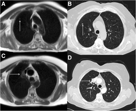
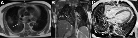
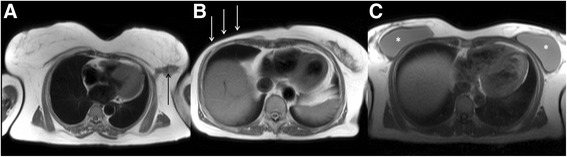
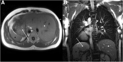
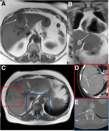
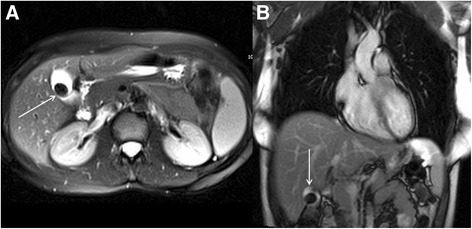
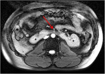
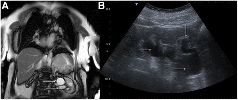
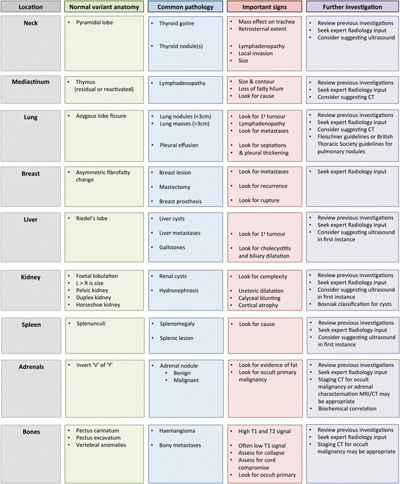
Cardiovascular magnetic resonance (CMR) is an established non-invasive technique to comprehensively assess cardiovascular structure and function in a variety of acquired and inherited cardiac conditions. A significant amount of the neck, thorax and upper abdomen are imaged at the time of routine clinical CMR, particularly in the initial multi-slice axial and coronal images. The discovery of unsuspected disease at the time of imaging has ethical, financial and medico-legal implications. Extra-cardiac findings at the time of CMR are common, can be important and can change clinical management. Certain patient groups undergoing CMR are at particular risk of important extra-cardiac findings as several of the cardiovascular risk factors for atherosclerosis are also risk factors for malignancy. Furthermore, the presence of certain extra-cardiac findings may contribute to the interpretation of the primary cardiac pathology as some cardiac conditions have multi-systemic extra-cardiac involvement. The aim of this review is to give an overview of the type of extra-cardiac findings that may become apparent on CMR, subdivided by anatomical location. We focus on normal variant anatomy that may mimic disease, common incidental extra-cardiac findings and important imaging signs that help distinguish sinister pathology from benign disease. We also aim to provide a framework to the approach and potential further diagnostic work-up of incidental extra-cardiac findings discovered at the time of CMR. However, it is beyond the scope of this review to discuss and determine the clinical significance of extracardiac findings at CMR.

Keywords: CMR; Cardiovascular Magnetic Resonance; Extra-cardiac findings; Extra-cardiac pathology; Incidental findings.


Figures

Fig. 1
Fig. 1
a to d Examples of the routine axial black blood images of the neck, thorax and upper abdomen acquired at the time of CMR (Cranial to Caudal). e to h Examples of the routine coronal bright blood images of the neck, thorax and upper abdomen acquired at the time of CMR (Anterior to Posterior)
Fig. 2
Fig. 2
CMR performed in a patient with bifasicular block and transient 2:1 second degree atrio-ventricular block. Short-axis LV cine images revealed global LV systolic impairment with an ejection fraction of 31 %. There is evidence of aorto-pulmonary (a), para-tracheal (a and b) and subcarinal (c) lymphadenopathy (solid white arrows) on axial black blood imaging. There is also subtle parenchymal lung infiltrate (solid red arrows) in the midzones (b and c) on axial black blood images. The short axis magnitude late gadolinium enhancement images (d) reveal septal mid-wall and patchy lateral wall replacement fibrosis (dashed white arrows). Together, the cardiac and extra-cardiac findings, in the clinical context are consistent with cardiac and pulmonary sarcoidosis
Fig. 3
Fig. 3
Axial black blood imaging of the neck (a) and coronal bright blood imaging (b) demonstrate homogeneous enlargement of the right lobe of thyroid gland (solid white arrows). There is local mass effect on the trachea with a degree of contralateral displacement and minor tracheal narrowing (*)
Fig. 4
Fig. 4
Axial black blood imaging of the thorax demonstrating a morphologically normal, ovoid left axillary lymph node (a) with fatty hilum (*). By convention, lymph node size (b) is measured in the short-axis (dashed black line)
Fig. 5
Fig. 5
Axial black blood imaging of the superior mediastinum in a 20 year old male patient who had recently suffered from a severe pneumonia complicated with myocarditis. The anterior mediastinal soft tissue (solid white arrows) detected at the time of CMR was felt too prominent to be dismissed as normal residual thymus and given the clinical history the diagnosis of thymic reactivation was made
Fig. 6
Fig. 6
Axial black blood imaging (a and b) and coronal bright blood imaging (c) demonstrating florid mediastinal adenopathy engulfing the trachea and proximal main bronchi (solid white arrows) detected incidentally at the time of CMR
Fig. 7
Fig. 7
Axial black blood imaging (a) and coronal bright blood imaging (b) acquired during a myocardial and hepatic iron loading protocol CMR in an individual with thalassaemia major. There are several prominent heterogeneous posterior mediastinal masses (solid white arrows) consistent with extramedullary haematopoeisis. The liver is uniformly low signal (white *), which is visual evidence of hepatic iron loading in the clinical context and the spleen is absent (black *) which may be the result of an auto-splenectomy
Fig. 8
Fig. 8
Axial black blood image of the superior mediastinum acquired at the time of CMR showing a linear high signal entity in the apical segment of the right upper lobe (solid white arrows) consistent with an incidental azygous lobe fissure
Fig. 9
Fig. 9
Axial black blood imaging (a) revealing an incidental 1.7 cm soft tissue density nodule in the right upper lobe (solid white arrows), which was subsequently confirmed with CT (b). Axial black blood imaging (c) demonstrating an incidental 3.1 cm soft tissue mass intimately related to the superior vena cava (solid white arrows), which was subsequently confirmed with CT (d)
Fig. 10
Fig. 10
Axial black blood imaging (a) and coronal bright blood imaging (b) demonstrating abnormal signal in the right sided pulmonary arterial system (solid white arrows). Magnitude late gadolinium enhancement 4-chamber image (c) showing signal void in the pulmonary arteries (solid white arrows) consistent with in situ thrombus from pulmonary embolic disease
Fig. 11
Fig. 11
Axial black blood imaging (a) showing an abnormal, spiculate soft tissue signal mass in the fatty left breast (solid black lesion) detected incidentally at the time of CMR. Axial black blood imaging (b) showing previous right-sided mastectomy (solid white arrows). Axial black blood imaging (c) showing bilateral breast prostheses (*)
Fig. 12
Fig. 12
Axial black blood imaging (a) and coronal bright blood imaging (b) demonstrating a left-sided liver (white *), right-sided spleen (black *) and right-sided stomach (solid white arrow). There is also evidence of a right-sided descending thoracic aorta on the axial and coronal images
Fig. 13
Fig. 13
Axial black blood imaging (a) and coronal bright blood imaging (b) showing a well-defined low and high signal entity respectively consistent with a simple liver cyst. Axial black blood imaging (c) showing ill-defined low signal entity in the right lobe of liver (red box) which demonstrated features consistent with a metastasis on contrast enhanced CT (d). There is also a low signal entity in the vertebral body (blue box) which was confirmed to represent a sclerotic metastasis on CT (e)
Fig. 14
Fig. 14
Axial (a) and coronal (b) bright blood imaging demonstrating a large low signal entity in a fluid-filled viscus in the right upper quadrant (solid white arrows), consistent with a large uncomplicated calcific gallstone
Fig. 15
Fig. 15
Axial bright blood imaging revealing a horseshoe kidney (*). The usual ascent of the kidneys out of the pelvis is impeded by the physical constraint of the inferior mesenteric artery (solid red arrow)
Fig. 16
Fig. 16
Coronal bright blood imaging (a) demonstrating a cystic entity at the left renal pelvis (solid white arrows). There is also a simple cortical renal cyst in the left lower pole. Correlative trans-abdominal ultrasound image (b) revealed the hypoechoic areas at the renal hilum are in continuity with a dilated proximal ureter and there is calyceal blunting (solid white arrows). These findings are consistent with hydronephrosis rather than para-pelvic cysts
Fig. 17
Fig. 17
Short-axis steady state free precession cine image (a) revealing a high signal cyst in the visualized left kidney (solid white arrow). On the corresponding magnitude late gadolinium enhancement image (b) there is no enhancement of the cyst (solid white arrow) but diffuse enhancement of the surrounding normal renal parenchyma. This is a reassuring feature of renal cystic lesions
Fig. 18
Fig. 18
Axial black blood imaging of the upper abdomen (a) revealing splenomegaly, with maximal bipolar diameter of the spleen of 17.1 cm (solid black arrows). This was detected incidentally at the time of CMR. Scrutinizing the other available extra-cardiac images (b) revealed extensive mediastinal lymphadenopathy (solid white arrows), suggesting the splenomegaly was due to lymphoma in this patient
Fig. 19
Fig. 19
Real-time first pass myocardial perfusion at peak stress following administration of intravenous adenosine (a) revealing no discernible enhancement of the visualized spleen (solid white arrow). Real-time first pass myocardial perfusion at rest in the same patient (b) revealing normal diffuse enhancement of the splenic tissue in the field of view (dashed white arrow)
Fig. 20
Fig. 20
Axial black blood imaging (a) and coronal bright blood imaging (b) revealing an incidental left adrenal nodule (solid black arrow) detected at the time of CMR
Fig. 21
Fig. 21
Axial black blood imaging (a) demonstrating abnormal soft tissue signal masses in the anticipated anatomical location of the adrenal glands (solid white arrows). These areas were confirmed to be highly metabolically active on metaiodobenzylguanidine (MIBG) study (b) confirming the diagnosis of bilateral phaeochromocytomas, which were the cause of this patient’s hypertension
Fig. 22
Fig. 22
In-phase MRI image (a) showing a right adrenal lesion (solid white arrow). There is signal drop out (dashed white arrow) on the out-of-phase MRI sequence (b), confirming the presence of intracellular lipid and making the diagnosis of a benign lipid-rich adenoma
Fig. 23
Fig. 23
Axial black blood lesion demonstrating a bony lesion in the right humeral head (solid white arrow)
Fig. 24
Fig. 24
4-Chamber steady state free precession cine showing mitral valve prolapse with central coaptation defect and a jet of mitral regurgitation (solid black arrows). There is a marked pectus exacavatum chest wall deformity (solid white arrow). The unifying diagnosis in this case was Marfan’s syndrome
Fig. 25
Fig. 25
Axial black blood imaging (a) and coronal bright blood imaging (b) showing a high signal entity within a thoracic vertebral body, with no involvement of the posterior elements (solid white arrows). The patient had previously undergone a thoracic CT (c) and this confirmed the classic appearance of a benign thoracic haemangioma with central ‘polka dot’ sign (white arrow)
Fig. 26
Fig. 26
Axial steady state free precession cine image (a) and main pulmonary artery long-axis stack steady state free precession cine image (b) revealing an abnormal mass within the spinal canal (solid white arrows) detected incidentally at the time of CMR
Fig. 27
Fig. 27
Normal variant anatomy, common pathology, important imaging signs and potential further investigations for extra-cardiac findings discovered at the time of CMR

References

    1. Bruder O, Wagner A, Lombardi M, Schwitter J, van Rossum A, Pilz G, et al. European Cardiovascular Magnetic Resonance (EuroCMR) registry--multi national results from 57 centers in 15 countries. J Cardiovasc Magn Reson. 2013;15:9. doi: 10.1186/1532-429X-15-9. - DOI - PMC - PubMed
    1. Hundley WG, Bluemke DA, Finn JP, Flamm SD, Fogel MA, Friedrich MG, et al. ACCF/ACR/AHA/NASCI/SCMR 2010 expert consensus document on cardiovascular magnetic resonance: a report of the American College of Cardiology Foundation Task Force on Expert Consensus Documents. J Am Coll Cardiol Elsevier Inc. 2010;55(23):2614–2662. doi: 10.1016/j.jacc.2009.11.011. - DOI - PMC - PubMed
    1. McMurray JJV, Adamopoulos S, Anker SD, Auricchio A, Böhm M, Dickstein K, et al. ESC Guidelines for the diagnosis and treatment of acute and chronic heart failure 2012: The Task Force for the Diagnosis and Treatment of Acute and Chronic Heart Failure 2012 of the European Society of Cardiology. Developed in collaboration with the Heart. Eur Heart J. 2012;33(14):1787–1847. doi: 10.1093/eurheartj/ehs104. - DOI - PubMed
    1. Kilner PJ, Geva T, Kaemmerer H, Trindade PT, Schwitter J, Webb GD. Recommendations for cardiovascular magnetic resonance in adults with congenital heart disease from the respective working groups of the European Society of Cardiology. Eur Heart J. 2010;31(7):794–805. doi: 10.1093/eurheartj/ehp586. - DOI - PMC - PubMed
    1. Dunet V, Schwitter J, Meuli R, Beigelman-Aubry C. Incidental extracardiac findings on cardiac MR: Systematic review and meta-analysis. J Magn Reson Imaging. 2015;23. - PubMed

Publication types