Case Reports
. 2016 May 9;10(1):113.
doi: 10.1186/s13256-016-0898-3.

Coexistence of lung cancer and immunoglobulin G4-related lung disease in a nodule: a case report

Affiliations
Case Reports

Coexistence of lung cancer and immunoglobulin G4-related lung disease in a nodule: a case report

Hiroki Tashiro et al. J Med Case Rep. .

Abstract

Background: Immunoglobulin G4-related disease is characterized by infiltration of immunoglobulin G4-positive plasmacytes in various organs. The radiological findings of lung involvement of immunoglobulin G4-related disease include hilar and mediastinal lymphadenopathies, thickness of bronchovascular bundles, peribronchovascular consolidation, and lung nodules. Although a pathological approach is needed to diagnose immunoglobulin G4-related disease, it is ordinarily diagnosed by biopsy from one lesion even if there are multiple lesions. We reported a rare case of the coexistence of immunoglobulin G4-related disease and lung cancer in the same lung nodule.

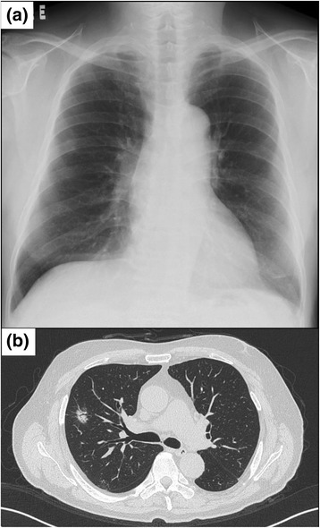
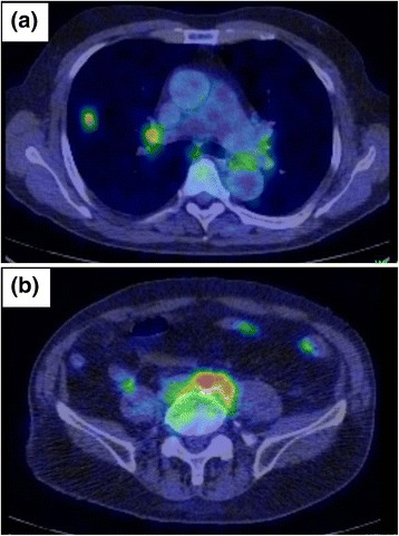
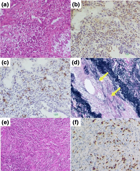
Case presentation: A 72-year-old Japanese man visited our hospital for evaluation of a nodular shadow in the middle lobe of his right lung that was seen on chest radiograph and computed tomography scan. An abdominal computed tomography scan showed a tumefactive lesion in his anterior sacral spine. Blood examinations revealed high serum immunoglobulin G4 concentration at 346 mg/dl, renal dysfunction, and anemia. He underwent right upper lobectomy and regional lymph node dissection. Pathologic findings of the lung nodule showed lepidic pattern adenocarcinoma with infiltration of immunoglobulin G4-positive plasma cells and obliterative phlebitis.

Conclusions: To date, there have been only few reports on the coexistence of immunoglobulin G4-related disease and lung cancer; here, we report such a rare case. Histologic examination should be considered in cases of suspicious immunoglobulin G4-related disease appearing in a lung nodule.

Keywords: IgG4-related disease; Lung cancer; Lung nodule.


Figures

Fig. 1
Fig. 1
The findings of a chest radiograph and computed tomography. a Chest radiograph shows a nodular shadow in the middle lobe of his right lung. b Chest computed tomography revealed a 20-mm spiculated part-solid nodule with pleural indentation in segment 3 on the right
Fig. 2
Fig. 2
Positron emission tomography-computed tomography examination in a 72-year-old man with coexisting lung cancer and immunoglobulin G4-related disease. There is high accumulation of 18F-fluorodeoxyglucose in his a lung nodule, hilar, and mediastinal lymph node and in his b retroperitoneum
Fig. 3
Fig. 3
Pathological findings of the resected lung nodule and hilar lymph node in a 72-year-old man with coexisting lung cancer and immunoglobulin G4-related disease. In the lung nodule, a lepidic pattern of adenocarcinoma coexisted with lymphocytes and plasma cells (hematoxylin and eosin, ×400). The infiltrating lymphocytes and plasma cells are positive for b immunoglobulin G and c immunoglobulin G4 staining (×400), with an immunoglobulin G4 to immunoglobulin G ratio of more than 40 %. d Obliterative phlebitis findings ("arrows") in the lung nodule (Elastica van Gieson, ×200). In the hilar lymph node, e the infiltration of plasma cells are seen with storiform fibrosis (×100) and f these cells were positive for immunoglobulin G4 staining (×400)

References

    1. Kamisawa T, Zen Y, Pillai S, Stone JH. IgG4-related disease. Lancet. 2015;385(9976):1460–71. doi: 10.1016/S0140-6736(14)60720-0. - DOI - PubMed
    1. Deshpande V, Zen Y, Chan JK, Yi EE, Sato Y, Yoshino T, et al. Consensus statement on the pathology of IgG4-related disease. Mod Pathol. 2012;25(9):1181–92. doi: 10.1038/modpathol.2012.72. - DOI - PubMed
    1. Campbell SN, Rubio E, Loschner AL. Clinical review of pulmonary manifestations of IgG4-related disease. Ann Am Thorac Soc. 2014;11(9):1466–75. doi: 10.1513/AnnalsATS.201403-128FR. - DOI - PubMed
    1. Matsui S, Hebisawa A, Sakai F, Yamamoto H, Terasaki Y, Kurihara Y, et al. Immunoglobulin G4-related lung disease: clinicoradiological and pathological features. Respirology. 2013;18(3):480–7. doi: 10.1111/resp.12016. - DOI - PubMed
    1. Fujinaga Y, Kadoya M, Kawa S, Hamano H, Ueda K, Momose M, et al. Characteristic findings in images of extra-pancreatic lesions associated with autoimmune pancreatitis. Eur J Radiol. 2010;76(2):228–38. doi: 10.1016/j.ejrad.2009.06.010. - DOI - PubMed

Publication types

MeSH terms

Substances