

doi: 10.1039/c6cc02764d.
Quantum dot-NanoLuc bioluminescence resonance energy transfer enables tumor imaging and lymph node mapping in vivo
Affiliations
- PMID: 27157466
- PMCID: PMC4912020
- DOI: 10.1039/c6cc02764d
Item in Clipboard
Quantum dot-NanoLuc bioluminescence resonance energy transfer enables tumor imaging and lymph node mapping in vivo
Chem Commun (Camb).
.
Abstract
A small luciferase protein (Nluc) was conjugated to QDs as a bioluminescence resonance energy transfer (BRET) pair. The conjugate showed 76% BRET efficiency and lymph node mapping was successfully performed. The cRGD peptide was conjugated to QD-Nluc for tumor targeting. The self-illuminating QD-Nluc showed excellent energy transfer in a living system and offered an optimal tumor-to-background ratio (>85).
Figures
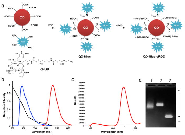
Characterization of bioluminescent-QD conjugates based on BRET. (a) Synthesis of QD-Nluc and QD-Nluc-cRGD conjugates. (b) Absorption and emission spectra of QD705 (λex = 465 nm), and bioluminescence of furimazine catalyzed by Nluc. (c) Bioluminescence emission spectrum of QD-Nluc in PBS. (d) Gel electrophoresis analysis of the conjugates; lane 1 is purified QD-Nluc conjugate; lane 2 is purified QD-Nluc-RGD conjugate, and lane 3 is unconjugated QD705.
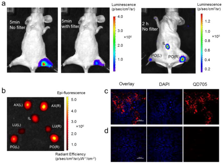
In vivo luminescence lymph node imaging. (a) Bioluminescence imaging of lymphatic basins in a mouse injected with QD-Nluc intradermally in a hind paw. (b) Fluorescence image of lymph nodes excised from the mouse injected with QD-Nluc in four paws (see Fig. S2); L, left; PO, popliteal lymph node; LU, lumbar lymph node; IL, iliac; AX, axillary lymph node; R, right. (c–d) Histological images of lymph node slices from a mouse in (a); (c) PO lymph node from injected mouse exhibited bright signal from QD705 (red) whereas no significant emission from QD705 was observed in PO lymph node from non-injected mouse (d). DAPI (blue) represents nuclease, scale bar: 20 μm.
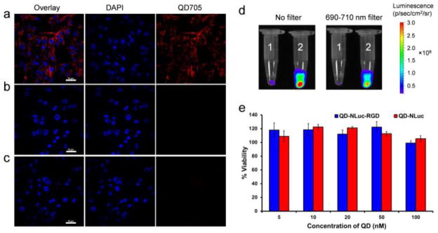
Fluorescence and luminescence imaging of U87MG cells with QD-Nluc-cRGD conjugate. (a–c) Confocal imaging; (a) cells were incubated with QD-Nluc-cRGD for 3 h, (b) cells were incubated with QD-Nluc for 3 h and (c) control cells without any conjugates, scale bar: 10 μm. (d) Luminescence images of labeled cells acquired without any filter (left) and with a filter (690–710 nm, right); cells were incubated with QD-Nluc (tube 1) and QD-Nluc-cRGD (tube 2). (e) Cell viability of U87MG cells evaluated by MTT assay in a dose-dependent manner of QD-Nluc and QD-Nluc-cRGD conjugates. Data represent mean ± s.d. (n = 4).
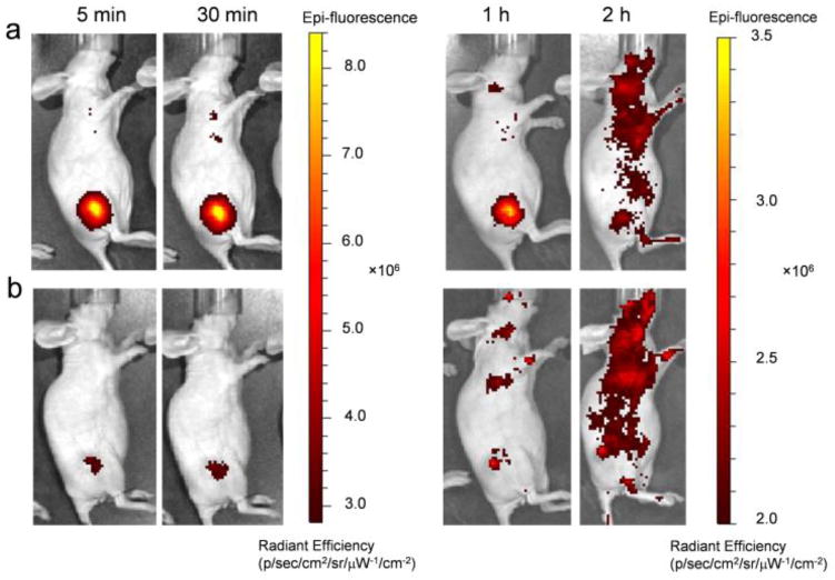
In vivo fluorescence imaging of U87MG tumor-bearing mice. (a,b) Time-dependent fluorescence imaging of U87MG tumor-bearing mouse intravenously injected with (a) QD-Nluc-cRGD or (b) QD-Nluc at 5 min, 30 min, 1 h and 2 h p.i..

Luminescence imaging of U87MG tumors in mice with QD-Nluc-cRGD. a,b) Time-dependent bioluminescence imaging of U87MG tumor-bearing mouse intravenously injected with QD-Nluc-cRGD (the same mouse as Fig. 4a) and single injection of furimazine substrate. Images in a) acquired without any emission filter with acquisition time: 1 s (5 min), 1 s (30 min), 1 s (1 h) and 10 s (2 h). Images in b) acquired with emission filter (690–710 nm) with acquisition time: 10 s (5 min), 10 s (30 min), 30 s (1 h) and 3 min (2 h).
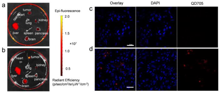
Ex vivo fluorescence imaging of organs and tumor tissues. a,b) Fluorescence images of organs excised from the mice in Fig. 4 injected with QD-Nluc (a) and QD-Nluc-cRGD (b). c,d) Histology of frozen U87MG tumors slices from a mouse injected with QD-Nluc (c) and QD-Nluc-cRGD (d). Scale bar: 10 μm.
Similar articles
-
Self-illuminating quantum dots for highly sensitive in vivo real-time luminescent mapping of sentinel lymph nodes.Int J Nanomedicine. 2012;7:3433-43. doi: 10.2147/IJN.S30709. Epub 2012 Jul 5. Int J Nanomedicine. 2012. PMID: 22848169 Free PMC article.
-
An enzymatically-sensitized sequential and concentric energy transfer relay self-assembled around semiconductor quantum dots.Nanoscale. 2015 May 7;7(17):7603-14. doi: 10.1039/c5nr00828j. Nanoscale. 2015. PMID: 25804284
-
Near infrared bioluminescence resonance energy transfer from firefly luciferase--quantum dot bionanoconjugates.Nanotechnology. 2014 Dec 12;25(49):495606. doi: 10.1088/0957-4484/25/49/495606. Epub 2014 Nov 21. Nanotechnology. 2014. PMID: 25414169
-
Integration of Nanomaterials and Bioluminescence Resonance Energy Transfer Techniques for Sensing Biomolecules.Biosensors (Basel). 2019 Mar 16;9(1):42. doi: 10.3390/bios9010042. Biosensors (Basel). 2019. PMID: 30884844 Free PMC article. Review.
-
NanoLuc: A Small Luciferase Is Brightening Up the Field of Bioluminescence.Bioconjug Chem. 2016 May 18;27(5):1175-1187. doi: 10.1021/acs.bioconjchem.6b00112. Epub 2016 Apr 19. Bioconjug Chem. 2016. PMID: 27045664 Free PMC article. Review.
Cited by
-
Chemiluminescence and Bioluminescence Imaging for Biosensing and Therapy: In Vitro and In Vivo Perspectives.Theranostics. 2019 May 31;9(14):4047-4065. doi: 10.7150/thno.33228. eCollection 2019. Theranostics. 2019. PMID: 31281531 Free PMC article. Review.
-
Creation of different bioluminescence resonance energy transfer based biosensors with high affinity to VEGF.PLoS One. 2020 Mar 26;15(3):e0230344. doi: 10.1371/journal.pone.0230344. eCollection 2020. PLoS One. 2020. PMID: 32214330 Free PMC article.
-
Coelenterazine-Dependent Luciferases as a Powerful Analytical Tool for Research and Biomedical Applications.Int J Mol Sci. 2020 Oct 10;21(20):7465. doi: 10.3390/ijms21207465. Int J Mol Sci. 2020. PMID: 33050422 Free PMC article. Review.
-
Current advances in the development of bioluminescent probes toward spatiotemporal trans-scale imaging.Biophys Physicobiol. 2024 Feb 2;21(Supplemental):e211004. doi: 10.2142/biophysico.bppb-v21.s004. eCollection 2024. Biophys Physicobiol. 2024. PMID: 39175853 Free PMC article. Review.
-
Nanobody-Nanoluciferase Fusion Protein-Enabled Immunoassay for Ochratoxin A in Coffee with Enhanced Specificity and Sensitivity.Toxins (Basel). 2022 Oct 19;14(10):713. doi: 10.3390/toxins14100713. Toxins (Basel). 2022. PMID: 36287981 Free PMC article.
References
Publication types
MeSH terms
Substances
Grants and funding
LinkOut - more resources
Full Text Sources
Other Literature Sources
Medical