

Prolonged maternal separation induces undernutrition and systemic inflammation with disrupted hippocampal development in mice
- PMID: 27157468
- PMCID: PMC4967409
- DOI: 10.1016/j.nut.2016.02.016
Prolonged maternal separation induces undernutrition and systemic inflammation with disrupted hippocampal development in mice
Abstract
Objective: Prolonged maternal separation (PMS) in the first 2 wk of life has been associated with poor growth with lasting effects in brain structure and function. This study aimed to investigate whether PMS-induced undernutrition could cause systemic inflammation and changes in nutrition-related hormonal levels, affecting hippocampal structure and neurotransmission in C57BL/6J suckling mice.
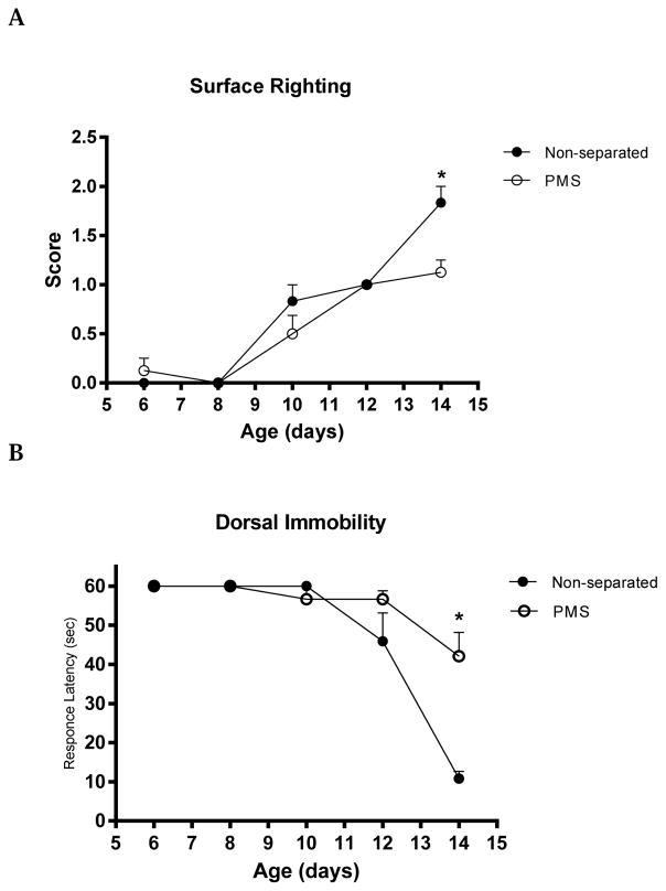
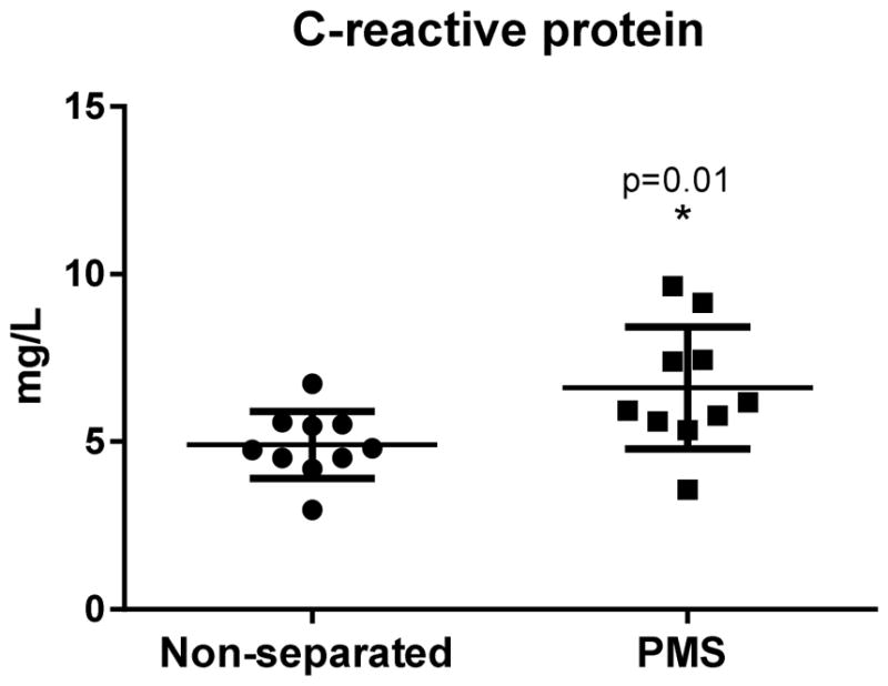
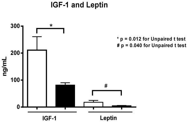
Methods: This study assessed mouse growth parameters coupled with insulin-like growth factor-1 (IGF-1) serum levels. In addition, leptin, adiponectin, and corticosterone serum levels were measured following PMS. Hippocampal stereology and the amino acid levels were also assessed. Furthermore, we measured myelin basic protein and synapthophysin (SYN) expression in the overall brain tissue and hippocampal SYN immunolabeling. For behavioral tests, we analyzed the ontogeny of selected neonatal reflexes. PMS was induced by separating half the pups in each litter from their lactating dams for defined periods each day (4 h on day 1, 8 h on day 2, and 12 h thereafter). A total of 67 suckling pups were used in this study.
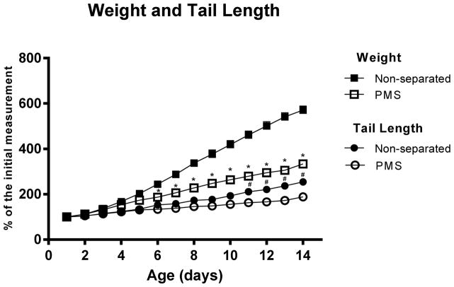
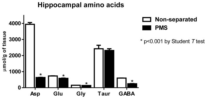
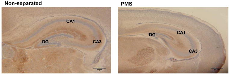
Results: PMS induced significant slowdown in weight gain and growth impairment. Significant reductions in serum leptin and IGF-1 levels were found following PMS. Total CA3 area and volume were reduced, specifically affecting the pyramidal layer in PMS mice. CA1 pyramidal layer area was also reduced. Overall hippocampal SYN immunolabeling was lower, especially in CA3 field and dentate gyrus. Furthermore, PMS reduced hippocampal aspartate, glutamate, and gamma-aminobutyric acid levels, as compared with unseparated controls.
Conclusion: These findings suggest that PMS causes significant growth deficits and alterations in hippocampal morphology and neurotransmission.
Keywords: Hippocampus; IGF-1; Inflammation; Leptin; Malnutrition; Maternal separation; Stereology.
Copyright © 2016 Elsevier Inc. All rights reserved.
Figures
Similar articles
-
Differential regulation of the insulin-like growth factors (IGF-I and -II) and IGF binding proteins during malnutrition in the neonatal rat.Endocrinology. 1991 Jul;129(1):149-57. doi: 10.1210/endo-129-1-149. Endocrinology. 1991. PMID: 1711459
-
Nutritional regulation of IGF-I expression during brain development in mice.Pediatr Res. 2001 Feb;49(2):197-202. doi: 10.1203/00006450-200102000-00011. Pediatr Res. 2001. PMID: 11158513
-
Postnatal maternal separation enhances tonic GABA current of cortical layer 5 pyramidal neurons in juvenile rats and promotes genesis of GABAergic neurons in neocortical molecular layer and subventricular zone in adult rats.Behav Brain Res. 2014 Mar 1;260:74-82. doi: 10.1016/j.bbr.2013.11.040. Epub 2013 Dec 1. Behav Brain Res. 2014. PMID: 24304720
-
Postnatal catch-up growth induced by growth hormone and insulin-like growth factor-I in rats with intrauterine growth retardation caused by maternal protein malnutrition.Pediatr Res. 1997 Sep;42(3):370-7. doi: 10.1203/00006450-199709000-00019. Pediatr Res. 1997. PMID: 9284279
-
Early-Life Stress Perturbs Key Cellular Programs in the Developing Mouse Hippocampus.Dev Neurosci. 2015;37(6):476-88. doi: 10.1159/000430861. Epub 2015 Jun 11. Dev Neurosci. 2015. PMID: 26068561 Free PMC article.
Cited by
-
Early-Life Stress Induces Depression-Like Behavior and Synaptic-Plasticity Changes in a Maternal Separation Rat Model: Gender Difference and Metabolomics Study.Front Pharmacol. 2020 Feb 26;11:102. doi: 10.3389/fphar.2020.00102. eCollection 2020. Front Pharmacol. 2020. PMID: 32174832 Free PMC article.
-
Maternal separation blunted spatial memory formation independent of peripheral and hippocampal insulin content in young adult male rats.PLoS One. 2018 Oct 17;13(10):e0204731. doi: 10.1371/journal.pone.0204731. eCollection 2018. PLoS One. 2018. PMID: 30332425 Free PMC article.
-
Inflammatory stimuli alter bone marrow composition and compromise bone health in the malnourished host.Front Immunol. 2022 Aug 2;13:846246. doi: 10.3389/fimmu.2022.846246. eCollection 2022. Front Immunol. 2022. PMID: 35983045 Free PMC article.
-
Understanding Role of Maternal Separation in Depression, Anxiety,and Pain Behaviour: A Mini Review of Preclinical Research With Focus on Neuroinflammatory Pathways.Int J Dev Neurosci. 2025 Feb;85(1):e70002. doi: 10.1002/jdn.70002. Int J Dev Neurosci. 2025. PMID: 39895419 Review.
-
Low maternal care enhances the skin barrier resistance of offspring in mice.PLoS One. 2019 Jul 11;14(7):e0219674. doi: 10.1371/journal.pone.0219674. eCollection 2019. PLoS One. 2019. PMID: 31295326 Free PMC article.
References
-
- Pollitt E. Developmental sequel from early nutritional deficiencies: conclusive and probability judgements. J Nutr. 2000;130:350S–353S. - PubMed
-
- Ivanovic DM, Leiva BP, Perez HT, Almagia AF, Toro TD, Urrutia M, et al. Nutritional status, brain development and scholastic achievement of Chilean high-school graduates from high and low intellectual quotient and socio-economic status. Br J Nutr. 2002;87:81–92. - PubMed
-
- Ivanovic DM, Leiva BP, Perez HT, Inzunza NB, Almagia AF, Toro TD, et al. Long-term effects of severe undernutrition during the first year of life on brain development and learning in Chilean high-school graduates. Nutrition. 2000;16:1056–1063. - PubMed
-
- Benitez-Bribiesca L, De lR-A I, Mansilla-Olivares A. Dendritic spine pathology in infants with severe protein-calorie malnutrition. Pediatrics. 1999;104:e21. - PubMed
-
- Beas-Zarate C, Ortuno-Sahagun D, Angel Meza AR, Feria-Velasco A. Effect of a corn diet during development on [3H]-spiperone binding in the brain of rats at the perinatal stage. Comp Biochem Physiol A Physiol. 1995;112:161–166. - PubMed
MeSH terms
Substances
Grants and funding
LinkOut - more resources
Full Text Sources
Other Literature Sources
Medical
Miscellaneous
