. 2016 Aug;24(7):1302-11.
doi: 10.1038/mt.2016.92. Epub 2016 May 9.

Analysis of the functional consequences of targeted exon deletion in COL7A1 reveals prospects for dystrophic epidermolysis bullosa therapy

Affiliations

Analysis of the functional consequences of targeted exon deletion in COL7A1 reveals prospects for dystrophic epidermolysis bullosa therapy

Olivier Bornert et al. Mol Ther. 2016 Aug.

Abstract

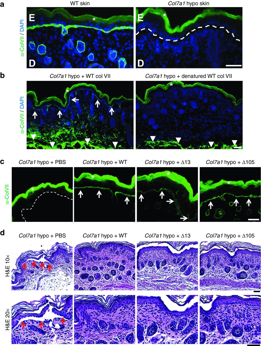
Genetically evoked deficiency of collagen VII causes dystrophic epidermolysis bullosa (DEB)-a debilitating disease characterized by chronic skin fragility and progressive fibrosis. Removal of exons carrying frame-disrupting mutations can reinstate protein expression in genetic diseases. The therapeutic potential of this approach is critically dependent on gene, protein, and disease intrinsic factors. Naturally occurring exon skipping in COL7A1, translating collagen VII, suggests that skipping of exons containing disease-causing mutations may be feasible for the treatment of DEB. However, despite a primarily in-frame arrangement of exons in the COL7A1 gene, no general conclusion of the aptitude of exon skipping for DEB can be drawn, since regulation of collagen VII functionality is complex involving folding, intra- and intermolecular interactions. To directly address this, we deleted two conceptually important exons located at both ends of COL7A1, exon 13, containing recurrent mutations, and exon 105, predicted to impact folding. The resulting recombinantly expressed proteins showed conserved functionality in biochemical and in vitro assays. Injected into DEB mice, the proteins promoted skin stability. By demonstrating functionality of internally deleted collagen VII variants, our study provides support of targeted exon deletion or skipping as a potential therapy to treat a large number of individuals with DEB.


Figures

Figure 1
Figure 1
In silico analysis of COL7A1 exon organization. The figure presents COL7A1 exons, organized as a jigsaw puzzle-like structure to illustrate which exons can be skipped without disrupting the open reading frame or changing more than one amino acid. Boxes with red line represent exons that cannot be skipped without evoking a reading frame shift. All such exons are located in noncollagenous domain 1 (NC1, in blue) and 2 (NC2, in green). The remaining exons correspond to the collagenous domain (in orange), which can be divided into two parts (P1 and P2) based on the presence of a noncollagenous hinge region in the middle of the protein. Localization of von Willebrand factor A (VWFA-1 and -2), FN type III and Kunitz inhibitor (Kunitz) domains are indicated as well.
Figure 2
Figure 2
Antisense oligonucleotides (AON) can induce skipping of exon 13 and 105 in dermal fibroblast. RT-PCR on RNA extracted from dermal fibroblast treated with 250 or 500 nmol/l of AONs designed against exon 13 (a) or 105 (c) shows efficient skipping of the respective exons. For both exons amplification of the region surrounding exon 13 (exon 12 to 14) or exon 105 (exon 102 to 106) reveals an additional lower band corresponding to an amplicon lacking exon 13 (144 bp) or 105 (81 pb), respectively. Sanger sequencing then confirms the precise skipping of exon 13 (b) or 105 (d). White asterisk indicates heteroduplex DNA.
Figure 3
Figure 3
Collagen VII variants can form stable triple helices. (a) The upper panel shows by RT-PCR on mRNA isolated from HEK cells expressing WT, Δ13, or Δ105 collagen VII that exon 13 or 105 are absent in the Δ13 and Δ105 construct carrying cells, respectively. The lower panel shows collagen VII immunostaining of HEK 293 cells expressing WT, Δ13 or Δ105 collagen VII. (b) Limited trypsin digestion at 30 °C with or without pre-boiling of the samples confirms that deletion of exon 13 or 105 in COL7A1 does not disturb the thermal stability of the collagen VII collagenous domain. (c) Detailed thermal stability analysis reveals that the Tm of all collagen VII variants (WT, Δ13, or Δ105) is approximately 42 °C.
Figure 4
Figure 4
Collagen VII variants retain ability to bind collagen IV. Variable concentrations of WT, denatured WT, Δ13, or Δ105 collagen VII were used (0.27 to 70 nmol/l) to analyze binding to 500 ng immobilized collagen IV. The data were normalized to the maximal signal recorded and were collected from three independent experiments. Heat denatured WT collagen VII shows a weak and nonsaturable binding and the collagen VII mutant G1776R shows a saturable interaction but with weaker affinity than for WT collagen VII. In contrast Δ13 and Δ105 collagen VII display similar Kd values as WT collagen VII indicating that binding to collagen IV is not disturbed by deletion of exon 13 or 105 from COL7A1.
Figure 5
Figure 5
Collagen VII variants support fibroblast adhesion and migration. Tissue culture plates were coated with bovine serum albumin, FN, WT, Δ13, or Δ105 collagen VII to study cell adhesion and migration. (a) Fibroblast adhesion assay. Two hours after seeding, fibroblasts were fixed and stained with crystal violet. After cell lysis, quantification was performed by recording the absorbance at 550 nm. Data are expressed as the percentage of adhesion. (b) Fibroblast wound healing assay. Dotted line represents the original wound edge. (c) Direct migration assay. Dotted line represents the original border. These results suggest that the lack of amino acids resulting from deletion of exon 13 or 105 does not interfere with the ability to support fibroblast adhesion and migration.
Figure 6
Figure 6
Collagen VII variants can be deposited at the dermal-epidermal junction in RDEB mice. (a) Skin tissue sections from WT and Col7a1 hypomorphic mice immunostained for collagen VII (green). While WT skin exhibits a strong signal for collagen VII at the dermal-epidermal junction zone, minimal collagen VII deposition is seen in Col7a1 hypomorphic mice (white dotted line). (b) Skin sections stained for human collagen VII (green) after intradermal injection of Col7a1 hypomorphic mice with 25 µg of WT collagen VII and heat denatured WT collagen VII. The staining reveals that only correctly folded collagen VII is able to translocate to the dermal-epidermal junction after injection (white arrows). Note that the injected collagen VII is clearly visible in deeper dermal areas in both skin sections (white arrowheads). (c) Skin sections one day after intradermal injection of Col7a1 hypomorphic mice with 10 µg of WT, Δ13, and Δ105 collagen VII stained for human collagen VII (green). The staining reveals that deletion of amino acids encoded by exon 13 or 105 does not affect the ability of collagen VII to translocate to the dermal-epidermal junction zone compared to WT collagen VII (white arrows). (d) H&E staining of skin sections as in (c). The histological analysis reveals that collagen VII variants lacking amino acids encoded by exon 13 or 105 promote dermal-epidermal stability equally well as WT collagen VII. While vehicle injected mice show areas of dermal-epidermal separation (red arrows), such separations are not visible in mice which had received 10 µg of WT, Δ13, or Δ105 collagen VII. Bar = 50 µm, D = dermis, E = epidermis, in a and b nuclei visualized by 4′,6-diamidino-2-phenylindole (blue). White asterisks indicate autofluorescence of the epidermis.

References

    1. Bruckner-Tuderman, L (2010). Dystrophic epidermolysis bullosa: pathogenesis and clinical features. Dermatol Clin 28: 107–114. - PubMed
    1. Mittapalli, VR, Madl, J, Löffek, S, Kiritsi, D, Kern, JS, Römer, W et al. (2016). Injury-driven stiffening of the dermis expedites skin carcinoma progression. Cancer Res 76: 940–951. - PubMed
    1. Vanden Oever, MJ and Tolar, J (2014). Advances in understanding and treating dystrophic epidermolysis bullosa. F1000Prime Rep 6: 35. - PMC - PubMed
    1. Goto, M, Sawamura, D, Ito, K, Abe, M, Nishie, W, Sakai, K et al. (2006). Fibroblasts show more potential as target cells than keratinocytes in COL7A1 gene therapy of dystrophic epidermolysis bullosa. J Invest Dermatol 126: 766–772. - PubMed
    1. Goto, M, Sawamura, D, Nishie, W, Sakai, K, McMillan, JR, Akiyama, M et al. (2006). Targeted skipping of a single exon harboring a premature termination codon mutation: implications and potential for gene correction therapy for selective dystrophic epidermolysis bullosa patients. J Invest Dermatol 126: 2614–2620. - PubMed

Publication types

MeSH terms

Substances