Current applications of transperineal ultrasound in gastroenterology
- PMID: 27158423
- PMCID: PMC4840194
- DOI: 10.4329/wjr.v8.i4.370
Current applications of transperineal ultrasound in gastroenterology
Abstract
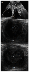
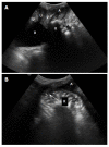
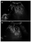
Transperineal ultrasound is an inexpensive, safe and painless technique that dynamically and non-invasively evaluates the anorectal area. It has multiple indications, mainly in urology, gynaecology, surgery and gastroenterology, with increased use in the last decade. It is performed with conventional probes, positioned directly above the anus, and may capture images of the anal canal, rectum, puborectalis muscle (posterior compartment), vagina, uterus, (central compartment), urethra and urinary bladder (anterior compartment). Evacuatory disorders and pelvic floor dysfunction, like rectoceles, enteroceles, rectoanal intussusception, pelvic floor dyssynergy can be diagnosed using this technique. It makes a dynamic evaluation of the interaction between pelvic viscera and pelvic floor musculature, with images obtained at rest, straining and sustained squeezing. This technique is an accurate examination for detecting, classifying and following of perianal inflammatory disease. It can also be used to sonographically guide drainage of deep pelvic abscesses, mainly in patients who cannot undergo conventional drainage. Transperineal ultrasound correctly evaluates sphincters in patients with fecal incontinence, postpartum and also following surgical repair of obstetric tears. There are also some studies referring to its role in anal stenosis, for the measurement of the anal cushions in haemorrhoids and in chronic anal pain.
Keywords: Fecal incontinence; Inflammatory perianal disease; Obstructed defecation; Posterior compartment; Transperineal ultrasound.
Figures


References
-
- Santoro GA, Wieczorek AP, Dietz HP, Mellgren A, Sultan AH, Shobeiri SA, Stankiewicz A, Bartram C. State of the art: an integrated approach to pelvic floor ultrasonography. Ultrasound Obstet Gynecol. 2011;37:381–396. - PubMed
-
- Valsky DV, Yagel S. Three-dimensional transperineal ultrasonography of the pelvic floor: improving visualization for new clinical applications and better functional assessment. J Ultrasound Med. 2007;26:1373–1387. - PubMed
-
- Oppenheimer DA, Carroll BA, Shochat SJ. Sonography of imperforate anus. Radiology. 1983;148:127–128. - PubMed
-
- Maconi G, Porro GB. Ultrasound of the gastrointestinal tract. 2nd ed. Springer, 2014
-
- Son JK, Taylor GA. Transperineal ultrasonography. Pediatr Radiol. 2014;44:193–201. - PubMed
Publication types
LinkOut - more resources
Full Text Sources
Other Literature Sources

