. 2016 Apr 10;8(1):e2016021.
doi: 10.4084/MJHID.2016.021. eCollection 2016.

Cutaneous Hyperpigmentation in Megaloblastic Anemia: a Five Year Retrospective Review

Affiliations

Cutaneous Hyperpigmentation in Megaloblastic Anemia: a Five Year Retrospective Review

Somanath Padhi et al. Mediterr J Hematol Infect Dis. .

Abstract

Background: Cutaneous hyperpigmentation is an often overlooked clinical sign in megaloblastic anemia (MA) which has been sporadically reported in the literature.

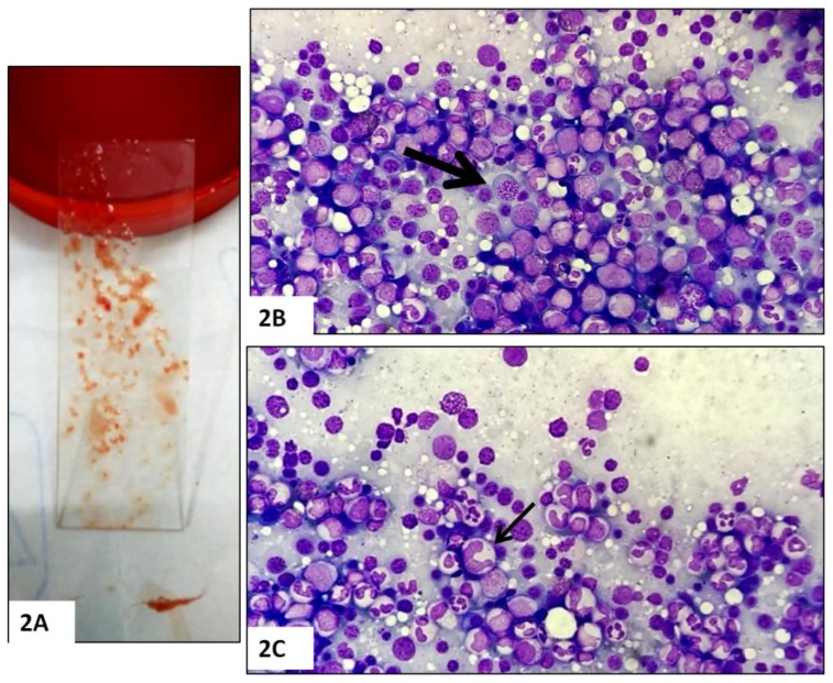
Methods: We describe the bone marrow (BM) changes and clinicolaboratory characteristics of 25 of 198 adult cases (>16 years) with cutaneous hyperpigmentation who underwent BM evaluation for cytopenia (s).

Results: Twenty-one of 25 cases (84%) had MA, while MA without hyperpigmentation occurred only in 12 of remainder 173 cases (P<0.001). Knuckle pad hyperpigmentation (KP) was noted in 16 (64%) cases; whereas 9 (36%) had diffuse brownish black discoloration (DP) of the palms and/or soles. Eighteen of 25 (72%) cases had pancytopenia (13 with KP) and 7 of 25 (28%) had bicytopenia (3 with KP). In addition, five cases (20%) presented with pyrexia. Of the 17 cases where data available, eleven were B12 deficient [<190 pg/ml; eight had severe deficiency (<100 pg/ml); ref.; 190-800pg/ml], while 4 had pure folate deficiency (< 4.0 ng/ml; ref.; 4-20ng/ml); and remainder 2 had combined B12 and folate deficiency. Compared to those with diffuse pigmentation; KP group had lower Hb (69.6 ± 24.2 vs. 86.3 ± 33.9 g/L), higher MCV (106.1 ±12.6 vs. 99.2 ± 7.6 fL), lower platelet count (50.9 ± 29.3 vs. 69.6 ± 36.5 × 10(9)/L), and lower median B12 [100.0 (30.0 - 822.0) vs. 316.0 (142.0 - 1617.3) pg/ml] (P>0.05). In six cases where follow-up data were available, there was a significant reversal of hyperpigmentation at 12 weeks following parenteral cobalamin therapy. In all five cases with pyrexia, fever subsided after 24 to 72 hours following administration of parenteral cobalamin therapy.

Conclusion: Cutaneous hyperpigmentation and cytopenia (s) are strongly associated with megaloblastic anemia. Knuckle pad hyperpigmentation is much more frequent than diffuse pigmentation of the palms and/or soles in such patents. A nonsignificant trend towards a greater degree of MA was found in cases with pigmentation of the knuckles.


Figures

Figure 1
Figure 1
Peculiar cutaneous hyperpigmentation from cases with megaloblastic anemia: typical diffuse, brownish-black discoloration of the palms (1A), knuckle pad hyperpigmentation in the dorsum of hands (1B) in a case of megaloblastic anemia (prior to therapy) in a 52 year old female who had gastric atrophy, proven on endoscopic biopsy (case no;18, Table 2). This patient had pancytopenia, macrocytosis (MCV; 116fL), and severe B12 deficiency (55pg/ml). Note the reversal of pigmentation (4A, 4B) in the same patient, 12 weeks after initiation of parenteral cyanocobalamin therapy. Diffuse brownish-black pigmentation over dorsal aspect of feet (1C) and dusky, brownish-black discoloration of palms with accentuation of palmar creases (1D) in a 52 year old vegetarian male with fever, jaundice, pancytopenia, macrocytosis (MCV; 115fL), and florid megaloblastic anemia proven on bone marrow examination (B12 and folate assay not done) (case no; 23, Table 2).
Figure 2
Figure 2. Bone marrow aspirate in megaloblastic anemia with cutaneous hyperpigmentation
Note the richly particulate bone marrow aspirate (2A) obtained during bone marrow procedure in cases with cutaneous hyperpigmentation and cytopenia (s). Bone marrow aspirate smears demonstrating erythroid hyperplasia and megaloblasts with sieve-like nuclear chromatin (2B, thick arrow) and giant, abnormal shaped stab forms (2C, thin arrow). These findings were consistent with a diagnosis of megaloblastic anemia (May Grunewald Giemsa, ×400).
Figure 3
Figure 3
Box plot diagram depicting the comparison of median (50th quartile, black horizontal line) and interquartile (25th to 75th) range of mean corpuscular volume (MCV) (A), Hb (B), Platelets (C), B12 (D), and folate (E) levels among two groups of pigmentation [knuckle pad (KP) vs diffuse type (DP)]. Note that the median and interquartile range of Hb, Platelets, and serum B12 were lower in the KP group than in the DP group; whereas the median and interquartile range of MCV were higher in KP group than the DP group. Also note that the group with DP has a wider B12 value compared to the KP group (D). The median value of serum folate was similar among two groups.
Figure 4
Figure 4
Reversal of hyperpigmentation in the patient of figure 1A and 1B, 12 weeks after initiation of parenteral cyanocobalamin therapy.
Figure 5
Figure 5
The postulated biochemical mechanism of hyperpigmentation in megaloblastic anemia., The 4 most accepted mechanisms involved are: 1) low methylcobalamin level in melanocytes leads to reduced level of reduced glutathione (GSSH); which in turn activates Tyrosinase enzyme in melanin synthesis pathway, 2) defective DNA synthesis activates Microphthalmia-associated transcription factor (MITF); which causes activation of both Tyrosinase and Tyrosinase related protein 1 and 2 (TRP 1and 2), 3) hyperhomocysteinemia leads to accumulation of cysteine leading to increased melanin synthesis, 4) defective melanin transfer from the melanocytes to adjacent megaloblastic keratinocytes. Increased angiogenesis secondary to upregulation of dermal vascular endothelial growth factor (VEGF) may also lead to increased pigmentation. Both histopathologic and ultrastructural studies have postulated that hyperpigmentation is due to increased number of basal melanocytes as well as increased melanosomes.

References

    1. Hoffbrand AV. Megaloblastic anemias. In: Fauci AS, Brunwald E, Kasper DL, et al., editors. Harrison’s Principles of Internal Medicine. 17th ed. New York: McGraw-Hill; 2008. pp. 643–651.
    1. Stabler SP. Vitamin B12 deficiency. N Eng J Med. 2013;368:149–160. doi: 10.1056/NEJMcp1113996. - DOI - PubMed
    1. Khanduri U, Sharma A. Megaloblastic anemia: prevalence and causative factors. Natl Med J India. 2007;20:172–175. - PubMed
    1. Arora S, Singh B, Gupta VK, Venkatesan M. Burden of vitamin B12 deficiency in urban population in Delhi, India: A hospital based study. Int J Pharma Biosci. 2011;2:B521–B528.
    1. Allen LH. How common is vitamin B12 deficiency? Am J Clin Nutr. 2009;89:693S–696S. doi: 10.3945/ajcn.2008.26947A. - DOI - PubMed