

Endoscopic and histological evaluations of a newly designed inguinal hernia mesh implant: Experimental studies on porcine animal model and human cadaver
- PMID: 27158482
- PMCID: PMC4846821
- DOI: 10.1016/j.amsu.2015.04.007
Endoscopic and histological evaluations of a newly designed inguinal hernia mesh implant: Experimental studies on porcine animal model and human cadaver
Abstract
Purpose: Conventional prostheses used for inguinal hernia repair are static and passive. This feasibility-study shows the features of a new 3D tension-free prosthesis in an experimental model.
Methods: This study was divided into two-phases: 1) aimed to test the physics intrinsic features and the anatomical adaptability of a new 3D designed mesh, and 2) aimed to evaluate the inflammatory reaction associated with different materials used. On phase-1 implantations were performed in pigs. During the first trial phase, the prostheses were also implanted on human cadavers. On phase-2, implantation was carried out on large swine. Follow-up was of 60-days, after which the animals were anaesthetized for laparoscopic assessment, and for sample collection of mesh implantation site for histological analysis.
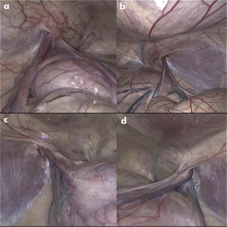
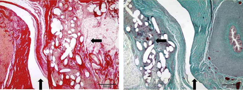
Results: All animals showed good 3D mesh tolerance, and the follow-up period was uneventful. The laparoscopy showed no inflammatory lesions on the internal surface of the peritoneum. Macroscopic observation of implantation site revealed some local fibrosis and reorganization of tissue, no signs of infection, and no changes on original implant positioning. Histological analysis on phase-1 showed in most sample segments the deferent duct maintaining its central position and surrounded by vascular and nervous structures. On phase-2 differences in inflammatory lesion score could be found between subjects.
Conclusions: This new 3D mesh can be placed appropriately and from this preliminary animal study no untoward complications were noted over a 60 day period.
Keywords: 3D mesh; Dynamic design; Experimental feasibility-study; Inguinal hernia.
Figures
References
-
- Rutkow I.M. Demographic and socioeconomic aspects of hernia repair in the United States in 2003. Surg Clin North Am. 2003;83:1045–1051. - PubMed
-
- Lichtenstein I., Shulman A. Ambulatory outpatient hernia surgery. Including a new concept, introducing tension-free repair. Int Surg. 1986;71:1–4. - PubMed
-
- Schwab R., Conze J., Willms A., Klinge U., Becker H.P., Schumpelick V. Management of recurrent inguinal hernia after previous mesh repair: a challenge. Chirurg. 2006;77:523–530. - PubMed
-
- Hernia E.U., Trialist Collaboration Mesh compared with nonmesh methods on open groin hernia repair. Systematic review of randomized controlled trial. Br J Surg. 2000;87:854–859. - PubMed
-
- Amid P.K. Lichtenstein tension-free hernioplasty: its inception, evolution, and principles. Hernia. 2004;8:1–7. - PubMed
LinkOut - more resources
Full Text Sources
Other Literature Sources
