Review
. 2016 Apr 7:2:16020.
doi: 10.1038/nrdp.2016.20.

Clostridium difficile infection

Affiliations
Review

Clostridium difficile infection

Wiep Klaas Smits et al. Nat Rev Dis Primers. .

Abstract

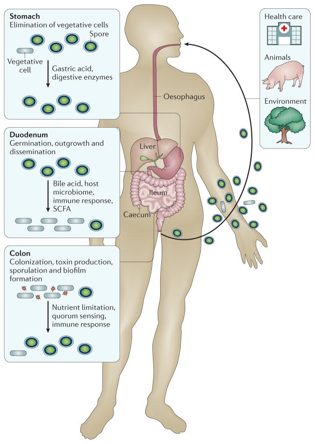
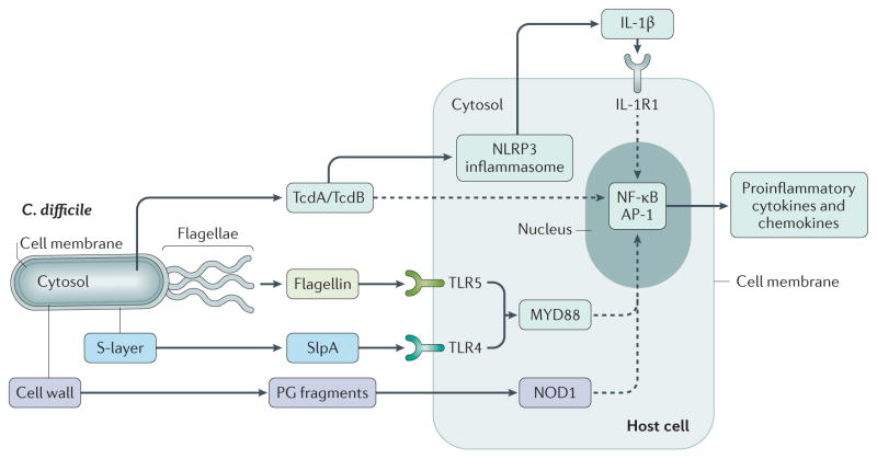
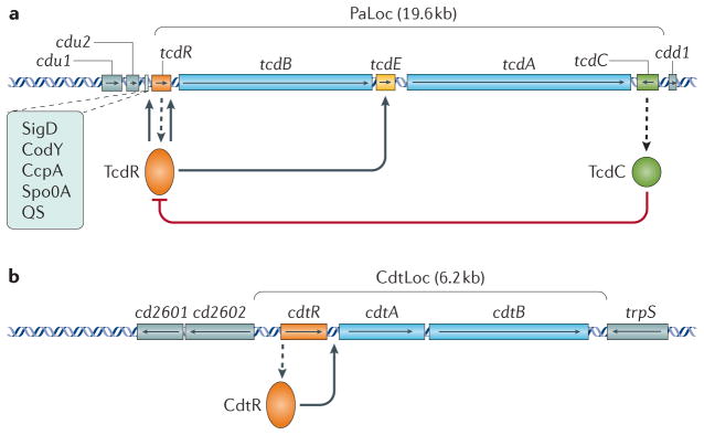
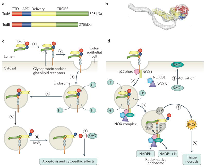
Infection of the colon with the Gram-positive bacterium Clostridium difficile is potentially life threatening, especially in elderly people and in patients who have dysbiosis of the gut microbiota following antimicrobial drug exposure. C. difficile is the leading cause of health-care-associated infective diarrhoea. The life cycle of C. difficile is influenced by antimicrobial agents, the host immune system, and the host microbiota and its associated metabolites. The primary mediators of inflammation in C. difficile infection (CDI) are large clostridial toxins, toxin A (TcdA) and toxin B (TcdB), and, in some bacterial strains, the binary toxin CDT. The toxins trigger a complex cascade of host cellular responses to cause diarrhoea, inflammation and tissue necrosis - the major symptoms of CDI. The factors responsible for the epidemic of some C. difficile strains are poorly understood. Recurrent infections are common and can be debilitating. Toxin detection for diagnosis is important for accurate epidemiological study, and for optimal management and prevention strategies. Infections are commonly treated with specific antimicrobial agents, but faecal microbiota transplants have shown promise for recurrent infections. Future biotherapies for C. difficile infections are likely to involve defined combinations of key gut microbiota.


Conflict of interest statement

Competing interests statement

W.K.S. has performed research for Cubist. D.L. has performed research for Immuron and Adenium Biotech. D.B.L. has performed research for MedImmune and Merck. M.H.W. has received consulting fees from Abbott, Actelion Pharmaceuticals, Astellas, AstraZeneca, Bayer, Cerexa, Cubist, Durata, The European Tissue Symposium, The Medicines Company, MedImmune, Merck, Motif Biosciences, Nabriva, Optimer, Paratek, Pfizer, Roche, Sanofi Pasteur, Seres Therapeutics, Summit Pharmaceuticals, and Synthetic Biologics. M.H.W. has also received lecture fees from Abbott, Alere, Astellas, AstraZeneca, Pfizer and Hoffmann La Roche, and received grant support from Abbott, Actelion, Astellas, bioMérieux, Cubist, Da Volterra, The European Tissue Symposium, Merck and Summit Pharmaceuticals. E.J.K. has performed research for Cubist, Novartis and Qiagen, and has participated in advisory forums of Astellas, Optimer, Actelion, Pfizer, Sanofi Pasteur and Seres Therapeutics. These companies had no role in the writing of this Primer.

Figures

Figure 1
Figure 1. Clostridium difficile
a | Typical image of Clostridium difficile colonies on a blood agar plate. b | Phase-contrast microscopy image of a C. difficile culture with vegetative cells (elongated rods), phase-dark spores (subterminal dark spots) and phase-bright spores (bright ellipsoids). Inset, Gram stain of culture. c | Scanning electron micrograph of C. difficile spores. d | Endoscopic picture of pseudomembranous colitis caused by C. difficile. Healthy colon tissue is pink, pseudomembranes resulting from C. difficile infection are yellow.
Figure 2
Figure 2. Stages of the Clostridium difficile life cycle in the human gastrointestinal tract
Three sources of infection (health care, animal and environmental) are indicated. A range of host factors influence the Clostridium difficile life cycle, as well as the relative numbers of spores and vegetative (metabolically active) cells in the gut. Note that passage through the stomach eliminates most vegetative cells (but spores survive), and spores germinate and grow out in the duodenum. In the caecum and colon, C. difficile starts producing spores again, and vegetative cells are excreted by the patient during infection. Toxin is produced in the colon. As C. difficile is an obligate anaerobic bacterium, transmission occurs primarily via spores. SCFA, short-chain fatty acid (such as butyrate).
Figure 3
Figure 3. Innate immune response of host cells to Clostridium difficile
Clostridium difficile elicits the innate immune response via at least four different effectors, leading to the induction of proinflammatory cytokines and chemokines via nuclear factor-κB (NF-κB) and transcription factor AP-1. The large clostridial toxins toxin A (TcdA) and TcdB act via NLRP3 (NOD-, LRR- and pyrin domain-containing 3) inflammasome-dependent and -independent pathways. Surface layer protein A (SlpA) and flagellin act via myeloid differentiation primary response 88 (MYD88)-dependent pathways through Toll-like receptor 4 (TLR4) and TLR5, respectively. The nucleotide-binding oligomerization domain-containing protein 1 (NOD1)-dependent pathway of induction most probably detects fragments of peptidoglycan (PG) derived from the cell wall of C. difficile. Dashed lines indicate indirect effects. IL-1R1, IL-1 receptor type 1.
Figure 4
Figure 4. Regulation of the Clostridium difficile toxins
a | Schematic representation of the pathogenicity locus (PaLoc) and the flanking regions with regulatory interactions of Clostridium difficile. Boxes with arrows indicate open reading frames with the direction of the arrows showing the direction of transcription. Toxin genes tcdA and tcdB are in blue, regulatory genes are in orange (positive) and green (negative); tcdE is in yellow and genes located outside the PaLoc are in grey. Dashed arrows indicate the production of protein from a gene transcript. Other regulators (Sigma D (SigD), the nutritional repressor CodY (known as GTP-sensing transcriptional pleiotropic repressor CodY), catabolite control protein A (CcpA), Stage 0 sporulation protein A (Spo0A) and quorum sensing (QS)) that affect toxin gene transcription (boxed) mostly act via expression of the tcdR gene. The TcdR protein is involved in the initiation of the production of toxin A (TcdA) and TcdB. b | Schematic representation of the binary toxin locus (CdtLoc) and flanking regions with regulatory interactions. Boxes with arrows indicate open reading frames with arrows showing the direction of transcription. The cdtA and cdtB toxin genes are in blue, the regulatory gene cdtR is in orange and genes located outside the CdtLoc are in grey.
Figure 5
Figure 5. Structure and function of the large clostridial toxins
a | Schematic of the toxin A (TcdA) and TcdB primary structures, highlighting the four functional domains: the glucosyltransferase domain (GTD; red), the autoprotease domain (APD; blue), the delivery domain (yellow) and the combined repetitive oligopeptides (CROPS) domain (green) that binds carbohydrates on the host cell surface to facilitate bacterial entry. b | Overlay of an electron microscopy reconstruction of the structure of TcdA with the X-ray crystal structure of TcdA lacking the CROPS domain (Protein Data Bank code: 4R04 ). Colour-coding reflects the domain structure in part a. c | The discrete structural and functional domains of the toxins contribute to a multi-step mechanism of intoxication. Toxins bind to one or more receptors (carbohydrate and/or protein) on the cell surface (step 1) and are internalized by receptor-mediated endocytosis (step 2). As the endosome matures, the V-ATPase contributes to a decrease in pH (step 3). The acidic pH causes a conformational change in the toxin delivery domain, resulting in pore formation (step 4) and the translocation of the APD and GTD into the cytosol (step 5). Inositol hexakisphosphate (InsP6) binds and activates the APD, resulting in the release of the GTD (step 6), which can inactivate RHO family proteins (step 7) to cause apoptosis and cytopathic ‘rounding’ effects. d | At concentrations >0.1 nM, TcdB can promote RAS-related C3 botulinum toxin substrate 1 (RAC1) activation (step 1) and complex formation between p22phox (also known as cytochrome b-245 light chain), NADPH oxidase 1 (NOX1), NADPH oxidase activator 1 (NOXA1), NADPH oxidase organizer 1 (NOXO1) and RAC1 on the endosomal membrane to form the NOX complex (step 2). The fully assembled NOX complex generates superoxide by transferring an electron from NADPH to molecular oxygen (step 3). Superoxide generation leads to the production of reactive oxygen species (ROS), which — at high levels — promote necrosis by causing mitochondrial damage, lipid peroxidation and protein oxidation (step 5).
Figure 6
Figure 6. Histopathology of Clostridium difficile infection in a mouse model
Histopathological analysis of haematoxylin and eosin-stained caecal and colonic tissues collected from mice infected with a wild-type PCR ribotype 027 strain (TcdA+ TcdB+), infected with an isogenic double tcdA and tcdB mutant (TcdA TcdB) or mock-infected with phosphate buffered saline (Mock). Note that both wild-type and double-mutant strains contain an intact binary toxin locus. Arrows indicate major histological differences; oedema and polymorphonuclear cell influx into the lamina propria (black arrows), erosion of crypts and goblet cell loss (yellow arrows) and hyperplasia (white arrows).
Figure 7
Figure 7. Mechanism of action of Clostridium difficile transferase (binary toxin)
Clostridium difficile transferase (CDT) is a binary toxin consisting of the CDTa ADP-ribosyltransferase (red) and the CDTb protein (yellow and green). The monomeric form of CDTb binds to the lipolysis-stimulated lipoprotein receptor (LSR), which is found in many tissues, including the gut. CDTb undergoes proteolytic activation and oligomerizes to form a heptameric prepore, which facilitates the binding of CDTa to the prepore–receptor complex. This complex enters cells by endocytosis and as the endosome matures, the V-ATPase contributes to a decrease in pH. The low pH of the endosome triggers pore formation and the translocation of CDTa into the cell. Once in the cytosol CDTa, ribosylates actin at arginine 177, resulting in a dual effect whereby G-actin polymerization is inhibited and F-actin depolymerization is favoured, which leads to the complete destruction of the actin cytoskeleton and, ultimately, cell death,. Inset, structure of CTDa (Protein Data Bank code: 2WN7 ).
Figure 8
Figure 8. Diagnosis and treatment options for Clostridium difficile infections
When a patient is suspected of having Clostridium difficile infection (CDI), the recommended option is to detect toxins of C. difficile in the stool. Several diagnostic algorithms have been condensed into this figure. Treatment options indicated here are based on reports by Leffler and Lamont, and Debast et al.. For moderate CDI, metronidazole is given orally, in severe cases intravenously. Hospitalization refers to admission as a result of CDI (not as a result of comorbidities; patients might already be hospitalized). Note that recurrence after clinical cure (resolution of symptoms) can be observed. Faecal microbiota transplant is an effective but non-standard form of treatment (shown by a dashed line). *Fidaxomicin is a treatment option if the risk of recurrence is high, but not for complicated CDI.
Figure 9
Figure 9. Faecal microbiota transplant
In faecal microbiota transplant (FMT), faecal material from a healthy donor is collected. The material is processed (blending, filtration) into pills or a solution. As part of this process, a check for the presence of pathogenic and multidrug-resistant organisms is performed. The processed material can be stored before (one-off) administration by nasoduodenal infusion, colonoscopic infusion or rectal enema for solution formulations or orally for pill formulations. Antibiotic treatment generally precedes the administration of the FMT to reduce Clostridium difficile levels. Alongside FMT, efforts are ongoing to standardize bacteriotherapy. On the basis of microbiome and metabolome analyses, signatures of resistance to colonization by C. difficile are identified. After collecting faecal material from healthy donors, species identified in these microbiome signatures or believed to be responsible for the metabolomic signature are cultured. Defined mixtures of these strains are tested for safety and ability to confer colonization resistance in preclinical trials and subsequently validated in clinical studies. Coloured bars indicate diversity of the microbiota, which is severely reduced in the patient with C. difficile infection (CDI) compared with the healthy subject.
Figure 10
Figure 10. Cost per case of Clostridium difficile infection
Data are from Ghantoji et al. (indicated with *) and Vonberg et al. (indicated with); surnames of first authors and years indicate the original studies described in these publications. Conversion between US$ and € is based on 2008 exchange rates. Note that several studies have estimated the cost of Clostridium difficile infections more recently,,. IBD, inflammatory bowel disease; ICU, intensive care unit.

References

    1. Hall IC, O’Toole E. Intestinal flora in new-born infants: with a description of a new pathogenic anaerobe, Bacillus difficilis. Am J Child Dis. 1935;49:390–402.
    1. Lyras D, et al. Toxin B is essential for virulence of Clostridium difficile. Nature. 2009;458:1176–1179. - PMC - PubMed
    1. Kuehne SA, et al. The role of toxin A and toxin B in Clostridium difficile infection. Nature. 2010;467:711–713. - PubMed
    1. Hafiz S, Oakley CL. Clostridium difficile: isolation and characteristics. J Med Microbiol. 1976;9:129–136. - PubMed
    1. Bartlett JG. Clostridium difficile: history of its role as an enteric pathogen and the current state of knowledge about the organism. Clin Infect Dis. 1994;18:S265–S272. An exceptional overview of the early experiments demonstrating the involvement of C. difficile in (antibiotic-associated) colitis. - PubMed

MeSH terms