

Neural Correlates of Antidepressant Treatment Response in Adolescents with Major Depressive Disorder
- PMID: 27159204
- PMCID: PMC5069704
- DOI: 10.1089/cap.2015.0232
Neural Correlates of Antidepressant Treatment Response in Adolescents with Major Depressive Disorder
Abstract
Objective: The neural changes underlying response to antidepressant treatment in adolescents are unknown. Identification of neural change correlates of treatment response could (1) aid in understanding mechanisms of depression and its treatment and (2) serve as target biomarkers for future research.
Method: Using functional magnetic resonance imaging, we examined changes in brain activation and functional connectivity in 13 unmedicated adolescents with major depressive disorder (MDD) before and after receiving treatment with a selective serotonin reuptake inhibitor medication for 8 weeks. Specifically, we examined brain activation during a negative emotion task and resting-state functional connectivity (RSFC), focusing on the amygdala to capture networks relevant to negative emotion. We conducted whole-brain analyses to identify how symptom improvement was related to change in brain activation during a negative emotion task or amygdala RSFC.
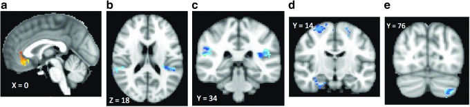
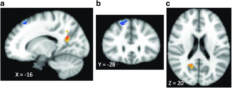
Results: After treatment, clinical improvement was associated with decreased task activation in rostral and subgenual anterior cingulate cortex and increased activation in bilateral insula, bilateral middle frontal cortices, right parahippocampus, and left cerebellum. Analysis of change in amygdala RSFC showed that treatment response was associated with increased amygdala RSFC with right frontal cortex, but decreased amygdala RSFC with right precuneus and right posterior cingulate cortex.
Conclusion: The findings represent a foothold for advancing understanding of pathophysiology of MDD in adolescents by revealing the critical neural circuitry changes that underlie a positive response to a standard treatment. Although preliminary, the present study provides a research platform for future work needed to confirm these biomarkers at a larger scale before using them in future target engagement studies of novel treatments.
Keywords: adolescent; antidepressants; depression; fMRI; treatment response.
Figures
References
-
- AACAP: Practice parameters for the assessment and treatment of children and adolescents with depressive disorders. J Am Acad Child Adolesc Psychiatry 37 (10 Supplement): 63S–83S, 1998 - PubMed
-
- Anand A, Yu Li, Wang Y, Wu J, Gao S, Bukhari L, Mathews VP, Kalnin A, Lowe MJ: Antidepressant effect on connectivity of the mood-regulating circuit: An fMRI study. Neuropsychopharmacology 30:1334–1344, 2005 - PubMed
-
- Beck AT, Steer RA: Beck Depression Inventory–Revised. San Antonio (TX), Harcourt Brace, 1996
Publication types
MeSH terms
Substances
Grants and funding
LinkOut - more resources
Full Text Sources
Other Literature Sources
Medical
