. 2016 Jun;79(6):1031-1037.
doi: 10.1002/ana.24650. Epub 2016 May 9.

Whole exome sequencing in patients with white matter abnormalities

Collaborators, Affiliations

Whole exome sequencing in patients with white matter abnormalities

Adeline Vanderver et al. Ann Neurol. 2016 Jun.

Abstract

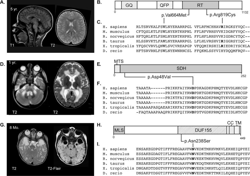
Here we report whole exome sequencing (WES) on a cohort of 71 patients with persistently unresolved white matter abnormalities with a suspected diagnosis of leukodystrophy or genetic leukoencephalopathy. WES analyses were performed on trio, or greater, family groups. Diagnostic pathogenic variants were identified in 35% (25 of 71) of patients. Potentially pathogenic variants were identified in clinically relevant genes in a further 7% (5 of 71) of cases, giving a total yield of clinical diagnoses in 42% of individuals. These findings provide evidence that WES can substantially decrease the number of unresolved white matter cases. Ann Neurol 2016;79:1031-1037.


Conflict of interest statement

Potential Conflicts of Interest

AV receives funding from Illumina, Inc., Gilead Sciences Inc., Eli Lilly & Co. and Shire Plc. AK, VR, ER, SC, TH, and RJT are employees of Illumina, Inc. The rest of the authors report no conflict of interest.

Figures

Figure 1
Figure 1. MRI and pathogenic variants for three cases
A) MRI of individual LD_0607.0 – a male of mixed European descent with a multisystem disorder characterized by elevated creatine kinase, recurrent infection with hypogammaglobulinemia, dyskeratosis congenita, and mild transaminase abnormalities. MRI revealed moderate cerebellar atrophy and diffuse multifocal white matter changes. Follow-up MRI one year later showed unchanged T2 hyperintensities. The variants found in TERT in this patient were classified as pathogenic per the ACMG guidelines and the diagnosis was supported by telomere length analysis (data not shown). B) Schematic of the TERT protein showing heterozygous variants identified in LD_0607.0. Predicted domains: GQ, GQ motif; QFP, QFP motif; RT, Reverse transcriptase domain. C) Clustalo alignment of vertebrate homologs of TERT showing conservation of mutated residues. D) MRI of individual LD_0756.0 – male of Turkish descent with motor delays were noted at birth who abruptly decompensated at 7 months of age, and a history of ataxia, hypotonia, and spasticity. MRI at 3 years and 6 months of age was significant for signal abnormality of the supratentorial white matter with sparing of the U fibers, a swollen appearance to the corpus callosum, involvement of the cerebellar white matter, and the brain stem. This pattern has been seen in previously published cases and supports the SDHB variant categorization as potentially pathogenic. E) Schematic of the SDHB protein showing a homozygous variant identified in LD_0756.0. Predicted domains: MTS, mitochondrial targeting signal; SDH, Succinate dehydrogenase domain. F) Clustalo alignment of vertebrate homologs of SDHB showing conservation of mutated residues. G) MRI of individual LD_0286.0B – male of mixed-European descent with leukoencephalopathy and a history of sensorineural hearing loss, developmental delay and febrile seizures. MRI is significant for bilateral temporal lobe cysts, small corpus callosum, and periatrial white matter abnormalities. Hearing loss and other clinical manifestations were consistent with the phenotype reported for mutations in RMND1, and the variant was classified as likely pathogenic based on supporting evidence. H) Schematic of the RMND1 protein showing heterozygous variants identified in LD_0286. Predicted domains: MLS, mitochondrial localization signal; DUF155, domain of unknown function 155; CC, coiled-coil domain; and TM, trans-membrane domain. I) Clustalo alignment of vertebrate homologs of RMND1 showing conservation of mutated residues.

References

    1. Vanderver A, Prust M, Tonduti D, et al. Case definition and classification of leukodystrophies and leukoencephalopathies. Mol Genet Metab. 2015 Apr;114(4):494–500. - PMC - PubMed
    1. Schiffmann R, van der Knaap MS. Invited article: an MRI-based approach to the diagnosis of white matter disorders. Neurology. 2009 Feb 24;72(8):750–9. - PMC - PubMed
    1. Vanderver A, Tonduti D, Schiffmann R, Schmidt J, Van der Knaap MS. Leukodystrophy Overview. In: Pagon RA, Adam MP, Bird TD, Dolan CR, Fong CT, Smith RJH, et al., editors. GeneReviews(R) Seattle (WA): University of Washington, Seattle; 2014. All rights reserved. - PubMed
    1. Parikh S, Bernard G, Leventer RJ, et al. A clinical approach to the diagnosis of patients with leukodystrophies and genetic leukoencephelopathies. Mol Genet Metab. 2015 Apr;114(4):501–15. - PMC - PubMed
    1. van der Knaap MS, Breiter SN, Naidu S, Hart AA, Valk J. Defining and categorizing leukoencephalopathies of unknown origin: MR imaging approach. Radiology. 1999 Oct;213(1):121–33. - PubMed

Publication types